Waking after surgery, you notice a patch of numb skin, a heavy-feeling hand, or a line of burning that runs down your leg. It’s unsettling, especially when no one has yet explained whether the sensation could be temporary. Postoperative nerve symptoms can have several causes, and the same symptom doesn’t always point to the same problem. Nerve injury after surgery is uncommon overall, but its frequency and consequences vary by operation, a patient’s existing nerve health, and how carefully symptoms are detected.
For patients whose symptoms persist after conservative care, mesenchymal stem cell (MSC) therapy at Dream Body Clinic offers a regenerative option aimed at supporting the body’s own repair processes rather than promising instant reversal.
Table of Contents
- What You May Be Feeling in the Hours After Surgery
- How Nerves Are Injured During Surgery
- Reading the Signs Sensory Motor and Pain Patterns
- When Testing Helps and When Waiting Harms
- Treatment Paths From Conservative Care to Surgical Repair
- How MSCs May Support Nerve Repair
- Recovery Timelines and Rehabilitation for International Patients
- Common Myths and Questions to Bring to Your Specialist
What You May Be Feeling in the Hours After Surgery
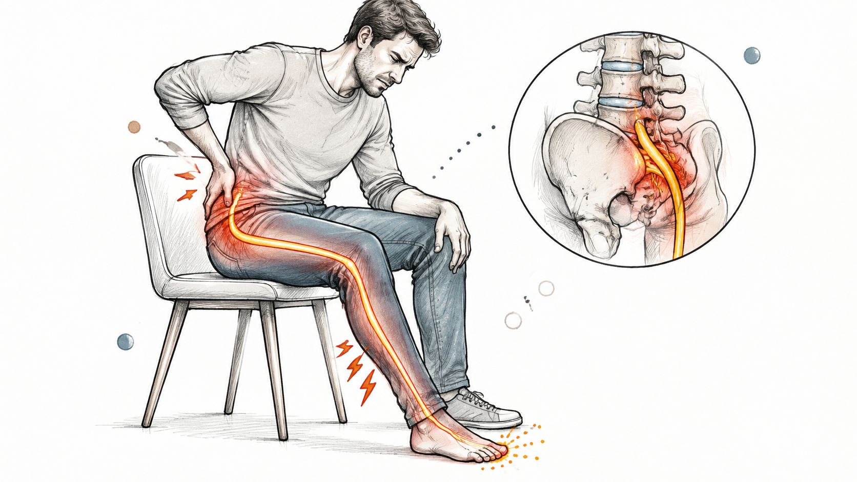
A patient wakes from an operation and finds that their fingers feel asleep. Another notices a burning strip along the thigh when they first try to stand. Someone else can feel touch but says their foot feels unusually weak. These sensations can be frightening, but they don’t all mean the same thing.
Nerves carry messages between the brain, spinal cord, and the rest of the body. Temporary irritation may cause tingling or numbness. When motor fibers are affected, weakness can appear. Pain may feel sharp, electric, or persistently sore. A sensory change and a loss of strength deserve different attention.
If symptoms remain after the early recovery period, some patients explore regenerative options such as the MSC protocols offered at Dream Body Clinic to help support the local environment around the injured nerve.
Why one overall rate can’t predict your case
Postoperative nerve injury is described as uncommon across surgery overall, but reported rates differ by procedure. One number can’t describe every operation or serve as a personal prognosis. Note what feels different, where you feel it, whether it’s changing, and whether you can move normally. Tell your care team about new symptoms, especially weakness or worsening pain.
That means one number can’t describe every operation or serve as a personal prognosis. (Review of postoperative nerve injury)
For now, note what feels different, where you feel it, whether it’s changing, and whether you can move normally. Tell your care team about new symptoms, especially weakness or worsening pain, rather than trying to decide on your own that they’re harmless.
How Nerves Are Injured During Surgery
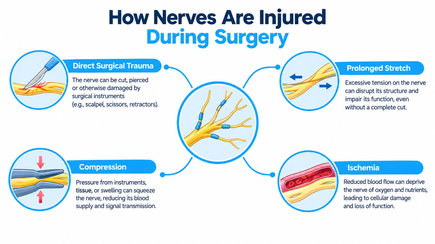
A nerve can stop transmitting normally if it is stretched, squeezed, deprived of blood supply, or physically cut. More than one of those stresses can act during the same operation. The distinction matters later: a conduction block from pressure may recover differently from an injury that damages axons.
Four pathways to a nerve problem
Direct trauma can occur when a nerve is cut, cauterized, retracted, or caught by a stitch. stretched or compressed by the position needed for surgery; the brachial plexus, which supplies the shoulder and arm, is vulnerable to excessive traction or pressure. In an anesthesia-claims analysis, the brachial plexus and ulnar nerve together accounted for about two-thirds of peripheral nerve injury claims, nearly half had no obvious single cause, and 91 percent were associated with anesthetic care judged appropriate (Anesthesia and surgery-related nerve injury review). Pressure can build after the operation as swelling or a hematoma crowds tissue around a nerve. Ischemia can interrupt signaling even when the nerve was never touched directly. Diabetes, existing neuropathy, or a spinal problem can lower a nerve’s tolerance for stress.
When residual inflammation or impaired signaling persists, the paracrine effects of MSCs used at Dream Body Clinic are intended to help modulate that environment and support resident repair cells.
Diabetes, existing neuropathy, or a spinal problem can lower a nerve’s tolerance for stress and change how symptoms are interpreted. Tell your clinician about prior numbness, weakness, or nerve pain. The operation may be where an older nerve problem first becomes noticeable.
Reading the Signs Sensory Motor and Pain Patterns
Describe what changed, not what you think the diagnosis is. “My fingertips are numb, but I can grip” gives a clinician different information from “I can feel my hand, but I can’t lift my wrist.” The pattern, location, and progression help guide the examination.
Sensory changes
Numbness, tingling, or burning are sensory symptoms. A clinician will consider the surgical site, the nerve’s path, positioning, an anesthetic block, and whether similar symptoms existed before surgery.
Weakness and pain
Difficulty lifting the front of the foot, raising an arm, or handling buttons may reflect a motor change. Pain can be burning, electric, sharp, or deep and aching. Persistent neuropathic pain is one reason some patients later consider MSC therapy to help calm inflammation and support the local repair environment around the affected nerve.
When Testing Helps and When Waiting Harms
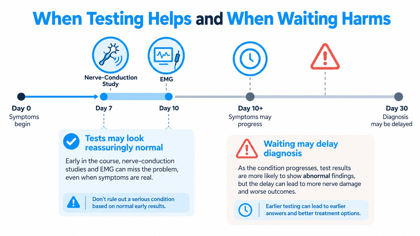
A nerve test can be useful and still be poorly timed. Nerve-conduction studies measure electrical signals travelling along nerves, and electromyography (EMG) records electrical activity inside muscles. A conduction study works like checking whether a cable still carries current, while EMG listens at the far end for signs the signal is no longer reaching the muscle. After an injury, the changes these tests detect take time to develop. During roughly the first 7–10 days, the results can be normal or only minimally abnormal despite a real deficit, as a review of electrodiagnostic timing explains.
Once the nature of the injury is clearer and the acute phase has passed, patients whose symptoms remain may discuss whether MSC therapy at Dream Body Clinic fits their situation.
Match the response to the symptoms
An initial study tends to be more informative around 2–4 weeks after injury, when denervation, axonal loss, and the likely site of damage become easier to identify. The same review notes that a repeat assessment roughly 4–6 weeks later can show whether reinnervation is beginning. Testing at this stage also helps separate a conduction block, which often recovers without intervention, from an axonal injury, which heals more slowly because the nerve fibre itself must regrow. Clinicians may add ultrasound or MRI when symptoms suggest compression or a structural cause (clinical review of postoperative nerve symptoms). If your symptoms followed a knee procedure, what a knee pop after ACL surgery can mean is a useful companion read.
Seek urgent assessment rather than waiting for a nerve test if weakness is worsening quickly, swelling is severe, circulation looks compromised, or a compressive hematoma is suspected. The same review emphasises that these situations may need imaging or treatment before an electrodiagnostic test can answer questions about the nerve.
For a stable sensory symptom, a clinician may choose observation with follow-up and testing at an appropriate point. Persistent motor loss or progressive symptoms call for early specialist review. A normal early study is not proof that nothing happened.
Practical rule: Don’t wait for a test appointment if movement is deteriorating or the limb looks or feels poorly perfused. Tell your surgical team what changed and when.
Treatment depends on where the nerve problem is, what caused it, which fibers are affected, and whether the deficit is improving. Options range from protection and activity adjustment to splinting, pain management, imaging, and, in selected severe cases, nerve surgery or reconstruction.
| Treatment Path | Best-Fit Injury Type | Primary Role in Recovery |
|---|---|---|
| Protection and activity adjustment | Symptoms that are mild or improving | Reduce avoidable strain and protect numb skin |
| Splinting and rehabilitation | Weakness that affects safe movement | Preserve useful motion and support function |
| Pain management | Persistent neuropathic pain | Ease symptoms while the underlying cause is evaluated |
| Imaging or targeted intervention | Suspected compression | Help locate the cause |
| Nerve surgery or reconstruction | Selected severe injuries | Relieve pressure or restore a pathway when appropriate |
| MSC therapy (Dream Body Clinic) | Persistent symptoms after conservative care | Support local repair signaling and inflammation modulation |
A clinician may recommend a brace, physical therapy, or skin checks where sensation is reduced. When residual pain or impaired function persists, some patients add regenerative support through the MSC protocols available at Dream Body Clinic.
How MSCs May Support Nerve Repair
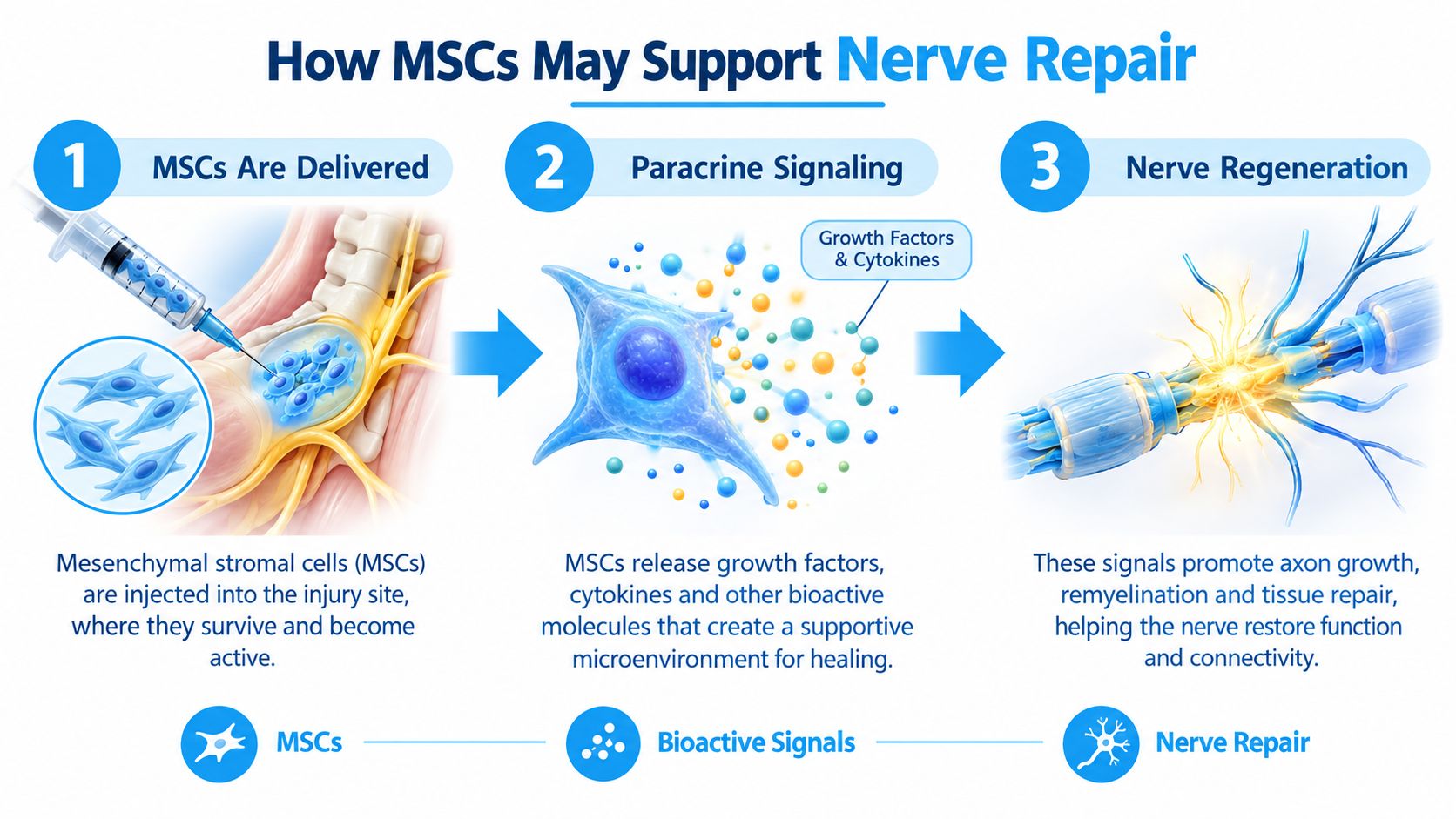
Mesenchymal stem cells do not transform into replacement nerve cells. Their proposed role is paracrine signaling: they release chemical messages that instruct nearby cells. Research reviews describe MSC effects as arising mainly from communication with resident repair cells such as Schwann cells, which form the channels along which regrowing axons extend in peripheral nerves.
MSCs can release neurotrophic factors including nerve growth factor (NGF), brain-derived neurotrophic factor (BDNF), glial-cell-line-derived neurotrophic factor (GDNF), and others associated with neuronal survival and axonal growth. They also release anti-inflammatory mediators that may help shift the local environment away from prolonged inflammation.
At Dream Body Clinic, allogeneic MSCs derived from Wharton’s jelly and placenta are expanded on site and can be delivered locally or systemically depending on the protocol chosen for the patient’s condition. The working model is messenger cells delivering repair instructions, with the patient’s own tissue doing the rebuilding. Signaling cannot rebuild a nerve that has been completely divided, so candidacy is assessed carefully.
For a plain-language explanation of related questions, see whether stem cell therapy may help peripheral neuropathy.
How MSCs May Support Nerve Repair
Mesenchymal stem cells do not transform into replacement nerve cells. Their proposed role is paracrine signaling: they release chemical messages that instruct nearby cells. Research reviews describe MSC effects as arising mainly from communication with resident repair cells such as Schwann cells, which form the channels along which regrowing axons extend in peripheral nerves.
MSCs can release neurotrophic factors including nerve growth factor (NGF), brain-derived neurotrophic factor (BDNF), glial-cell-line-derived neurotrophic factor (GDNF), and others associated with neuronal survival and axonal growth. They also release anti-inflammatory mediators that may help shift the local environment away from prolonged inflammation.
At Dream Body Clinic, allogeneic MSCs derived from Wharton’s jelly and placenta are expanded on site and can be delivered locally or systemically depending on the protocol chosen for the patient’s condition. The working model is messenger cells delivering repair instructions, with the patient’s own tissue doing the rebuilding. Signaling cannot rebuild a nerve that has been completely divided, so candidacy is assessed carefully.
For a plain-language explanation of related questions, see whether stem cell therapy may help peripheral neuropathy.
Signals, not replacement nerves
MSCs can release growth factors, cytokines, and extracellular vesicles. These signals may reach neurons, Schwann cells, glial cells, immune cells, and cells that support blood vessels. Schwann cells matter here because they form the channels along which regrowing axons extend in peripheral nerves. Research reviews describe MSC effects as arising mainly from communication with these resident repair cells, with the MSCs shifting the environment around the injury toward repair. (Review of MSCs in peripheral nerve repair)
This framing helps explain where researchers see the most plausible use. A signaling treatment needs repair machinery to signal to, so interest centers on injuries where the nerve’s supporting structure survives and cells like Schwann cells remain able to respond.
The same review describing paracrine signaling lists the neurotrophic factors MSCs release: nerve growth factor (NGF), brain-derived neurotrophic factor (BDNF), glial-cell-line-derived neurotrophic factor (GDNF), fibroblast growth factor 2 (FGF-2), ciliary neurotrophic factor (CNTF), insulin-like growth factor 1 (IGF-1), and hepatocyte growth factor (HGF). Each has been associated with neuronal survival, axonal growth, or a local environment more favorable to repair. A separate line of research describes MSC-linked anti-inflammatory mediators, including interleukin-1 receptor antagonist (IL-1Ra), tumor-necrosis-factor-stimulated gene-6 (TSG-6), prostaglandin E2 (PGE2), and interleukin-10 (IL-10), along with extracellular vesicles that may carry signals influencing inflammation and repair. (Review of MSC anti-inflammatory signaling)
This mechanism has limits worth stating plainly. A signal that supports repair in a laboratory model does not automatically restore function in a person with a particular injury, and signaling cannot rebuild a nerve that has been completely divided. Interest in regenerative care should never delay assessment of a worsening deficit, since some surgical repairs have limited time windows. If you are weighing this kind of treatment, ask the clinic how it describes the mechanism, what evidence supports the proposed use, and how your progress would be measured over time. For a plain-language explanation of how these questions apply in practice, see whether stem cell therapy may help peripheral neuropathy.
The working model is messenger cells delivering repair instructions, with your own tissue doing the rebuilding.
Recovery Timelines and Rehabilitation for International Patients
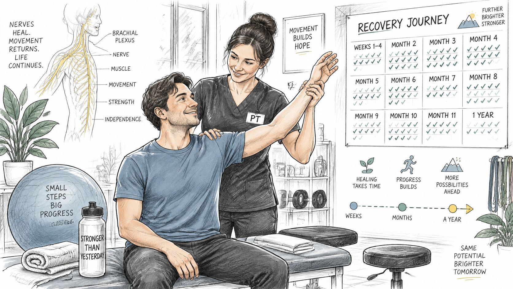
Recovery time follows the type of injury. Neurapraxia often improves as swelling settles. Axonotmesis requires slower regrowth along the preserved sheath. Neurotmesis usually needs surgical repair for meaningful recovery. Rehabilitation protects what you still have while the diagnosis is clarified.
Cross-border care adds the need for clear information sharing. Dream Body Clinic provides remote follow-up for international patients, typically at intervals that allow progress to be tracked. Arrange for operative notes, examination findings, and test results to be available so the team can determine whether an MSC protocol is appropriate alongside continued rehabilitation at home.
ommon Myths and Questions to Bring to Your Specialist
Myth: numbness automatically means permanent damage. A sensory change can come from swelling, temporary conduction block, or a deeper axonal injury. The pattern and progression matter more than the presence of numbness alone.
Myth: a normal early nerve test settles the question. Testing too soon can look reassuring while injured fibers are still changing.
Myth: MSCs become replacement nerves. The supported rationale is paracrine signaling. They do not grow into a new nerve cable.
Myth: one visit should reverse a chronic deficit. A credible plan states the target, the likely ceiling, and how progress will be measured.
Questions worth asking include which nerve or region fits the symptoms, what change would count as recovery, which symptoms mean urgent review, and—if regenerative care is proposed—what it is expected to influence and what it cannot repair.
Dream Body Clinic offers regenerative medicine services, including approaches used for nerve-related symptoms, with clinical assessment and remote follow-up for international patients. If you are exploring whether MSC therapy may fit your situation, visit Dream Body Clinic or call (888) 704-3977 to discuss your records and current symptoms.
7 Ways Mesenchymal Stem Cell Therapy at Dream Body Clinic Can Support Nerve Recovery
- Paracrine signaling — MSCs release neurotrophic factors and anti-inflammatory mediators that support the local repair environment.
- Support for Schwann cells and resident repair cells — Helps create conditions more favorable for axonal regrowth in peripheral nerves.
- Inflammation modulation — May help reduce the prolonged inflammatory response that can impede recovery.
- No harvest required — Allogeneic MSCs from Wharton’s jelly and placenta; no bone marrow or fat draw.
- Flexible delivery — Local or systemic protocols tailored after review of imaging and clinical findings.
- On-site expansion — Cells processed in the clinic’s ISO-rated lab and administered fresh.
- International patient support — Assessment, treatment in the Puerto Vallarta area, and remote follow-up for patients traveling from abroad.