In a study of 119 older adults, ligamentum flavum thickness didn't consistently distinguish symptomatic spinal stenosis from other groups, and among participants with clinical stenosis, it didn't track walking performance, disability, or pain scores, according to the Michigan Spinal Stenosis Study. That finding changes how an MRI report should be read: a thick ligament can contribute to nerve compression, but its measurement alone can't explain your symptoms or choose your treatment.
Ligamentum flavum thickening means that a ligament along the back of the spinal canal has become bulkier. The useful questions are where it's thickened, what space remains for the nerves, and whether the examination matches the scan. For patients considering treatment, including regenerative care, that distinction prevents an imaging finding from becoming an unsupported diagnosis.
Table of Contents
- Ligamentum Flavum Thickness Values Compared
- How to Read Thickness on an MRI Report
- Why the Ligament Thickens With Age and Stress
- How Thickening Narrows the Canal and Causes Symptoms
- Treatment Pathways From Conservative Care to Decompression
- Where Regenerative Stem Cell Therapy Fits
- Confirm Your Imaging Before Booking a Treatment
Ligamentum Flavum Thickness Values Compared
Ligamentum flavum thickness reference values
| Measure | Group or context | Value |
|---|---|---|
| Half-length thickness | Healthy lumbar spine | 3.1 ± 0.8 mm |
| Half-length thickness | Degenerative group | 4.9 ± 1.0 mm |
| Half-length thickness | Stenotic group | 5.3 ± 0.7 mm |
| Mean thickness | Stenosis patients vs controls | 4.24 ± 0.97 mm vs 3.61 ± 0.72 mm |
| Mean area | Stenosis patients vs controls | 132.69 ± 32.68 mm² vs 96.56 ± 30.74 mm² |
| Mean thickness | Mild vs severe claudication | 4.99 ± 1.20 mm vs 6.09 ± 1.39 mm |
| Hypertrophy at least 4 mm | Surgical cohort 2013–2017 vs 2018–2023 | 24.5% (24/98) vs 44.2% (46/104) |
| Hypertrophy greater than 4 mm | 2025 two study groups | 55.8% (91/163) and 89.5% (17/19) |
| Mean area and cutoff | Cervical stenosis vs controls, 2023 | 45.34 ± 9.09 mm² vs 25.24 ± 6.43 mm²; cutoff 31.66 mm² (92.8% sensitivity, 88.4% specificity) |
The half-length measurements and mild-versus-severe claudication values are reported in the Asian Spine Journal discussion of ligamentum flavum thickness. These comparisons support an association between greater thickness and stenotic disease. Group averages are not individual diagnostic boundaries, and the rows mix lumbar and cervical data with different definitions.
In the comparison of 166 stenosis patients and 167 controls, both mean thickness and mean area were greater in the stenosis group, with P < 0.001, as described in the lumbar ligamentum flavum area study. Area describes the ligament's cross-sectional bulk, while thickness describes a linear measurement. Neither identifies, without the surrounding images, exactly which nerve is compressed.
Read the population before interpreting the number
The surgical-cohort row describes people already selected for lumbar-stenosis surgery, not the general population. That 2025 retrospective cohort used an at-least-4-mm definition, found the rates shown above, and reported higher hypertrophy rates in patients aged 60 or older, with p = 0.004.
The other 2025 Asian Spine Journal series used greater than 4 mm, reporting the different rates shown in its study groups. The boundary differs: “at least” includes the boundary value, while “greater than” excludes it. Those percentages should not be treated as interchangeable prevalence estimates.
The cervical row concerns area, not a lumbar-style thickness threshold. The 2023 Medicine cervical-area study reported the listed group means and diagnostic performance; it does not establish a cervical thickness cutoff of 3 mm.
Practical rule: Compare the spinal region, level, measurement method, and patient group before comparing your result with a published value.
A measurement resembling the severe-claudication group should prompt careful inspection for thecal-sac or nerve-root compression. It does not establish severe symptoms by itself. No lumbar millimeter cutoff is universal, and neither side should be interpreted without the space surrounding it.
How to Read Thickness on an MRI Report
A useful MRI review connects the measurement to anatomy rather than highlighting the largest number. Have the full images available, not just the report's conclusion, so the clinician can check where and how the ligament was measured.
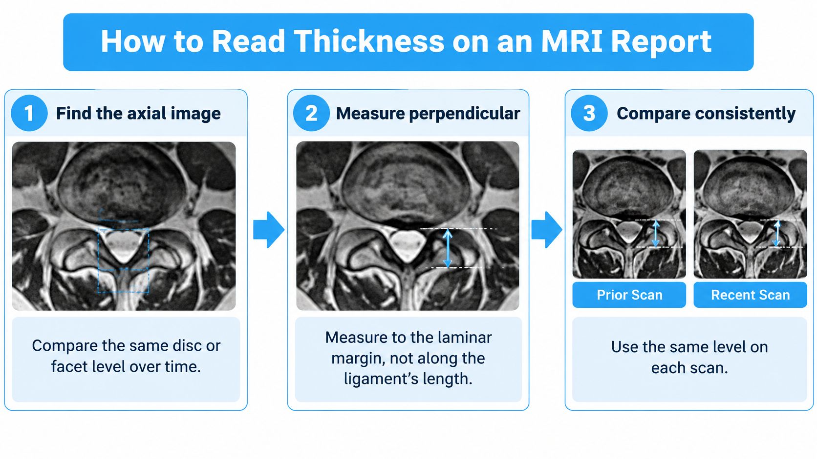
The measurement sequence is illustrated below.
Follow a consistent measurement sequence
Find the axial image at the relevant level. Axial images show a cross-section of the canal and its surrounding structures. Compare the same disc or facet-joint level across scans, because a measurement taken elsewhere can create the appearance of change.
Check the measurement direction. Thickness should be measured perpendicular to the laminar margin, rather than along the ligament's length. An oblique line can make the tissue appear thicker without showing a true increase in bulk.
Record the right and left sides separately. In an MRI comparison published in Spine, average thickness at L3–L4 was 2.90 mm on the right versus 2.76 mm on the left, and at L5–S1 it was 3.42 mm versus 3.22 mm. Side-specific reporting matters when symptoms and lateral-recess narrowing are asymmetric.
Inspect the remaining canal space. Ask the reviewer to consider disc protrusion, facet hypertrophy, epidural fat, and the outline of the thecal sac beside the ligament. A thickness value describes one structure, whereas compression reflects the combined arrangement of structures.
Identify what kind of tissue change is present. Look for descriptions of calcification, ossification, a cyst, hematoma, or another focal lesion. These terms aren't interchangeable with ordinary degenerative thickening, and they can change the diagnostic pathway.
Clarify whether CT or additional imaging is needed. MRI may show posterior canal narrowing without reliably separating soft-tissue hypertrophy from mineralization; the Asian Spine Journal review of ligamentum flavum lesions explains the importance of characterizing these alternatives. CT can help identify calcification or ossification, while a radiologist may recommend contrast imaging when a lesion needs further assessment.
Ask for a tissue description, not just a dimension
A report that mentions a focal mass near the ligament needs a different conversation from one describing broad degenerative thickening. A rare ligamentum flavum hematoma can resemble a disc herniation, tumor, or other epidural lesion, and its MRI appearance may change as blood products evolve.
The actionable conclusion should therefore identify the tissue abnormality, its location, and its effect on neural structures. A number without those details leaves the main clinical question unanswered.
Why the Ligament Thickens With Age and Stress
Consider a spinal segment where the disc and facet joints have developed degenerative changes. Repeated loading acts on the ligament beside them, and its cells respond by remodeling the tissue. The result isn't necessarily just a normal ligament folding inward.
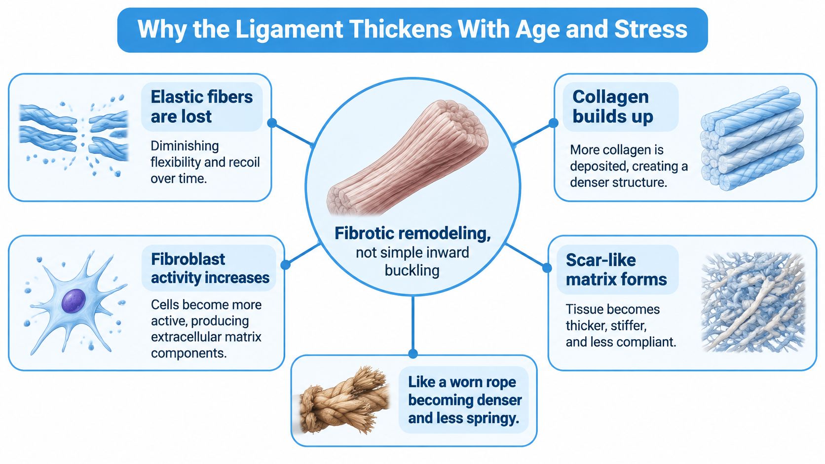
A useful analogy is a stretchy elastic band gradually becoming more like thick leather. Both occupy space, but the leather is less springy and less able to recover its original shape. In ligamentum flavum thickening, progressive loss of elastic fibers accompanies collagen accumulation and scar-like tissue formation, as described in the FASEB Journal review of ligamentum flavum remodeling.
The tissue changes are summarized in this diagram.
From mechanical loading to fibrotic tissue
Fibroblasts maintain the tissue's supporting framework. With chronic mechanical stress, segmental instability, inflammation, and oxidative stress, their activity can shift toward a more fibrotic pattern.
Signals including TNF-alpha, IL-1 beta, IL-6, and TGF-beta1 help drive this response. Some encourage fibroblasts to behave more like myofibroblasts, cells associated with tissue contraction and scar formation. In some circumstances, remodeling also includes bone-forming changes or ossification.
This biology matters because buckling and true thickening aren't the same process. A ligament can fold inward when its mechanical environment changes, but fibrotic enlargement involves changes within the tissue itself. Both can contribute to narrowing, and they may coexist.
Age is a context, not a countdown
The review describes an MRI study of 648 people in which thickening at L4–L5 had already begun at ages 30–39, with greater age-related progression at L3–L4 and L4–L5 than at upper or lumbosacral levels. Those observations describe a population pattern, not a prediction that a particular younger patient will develop symptomatic stenosis.
Disc and facet degeneration also frequently coexist with ligament remodeling. Patients exploring these overlapping findings can use the clinic's explanation of degenerative disc disease and spondylosis to distinguish the terminology.
A ligament can be biologically abnormal without being the main reason a patient hurts.
The practical interpretation is additive: the disc, facets, and ligament can each reduce available space. The clinician must judge the combined narrowing and neurologic findings, rather than assigning pain to whichever structure sounds most abnormal in the report.
How Thickening Narrows the Canal and Causes Symptoms
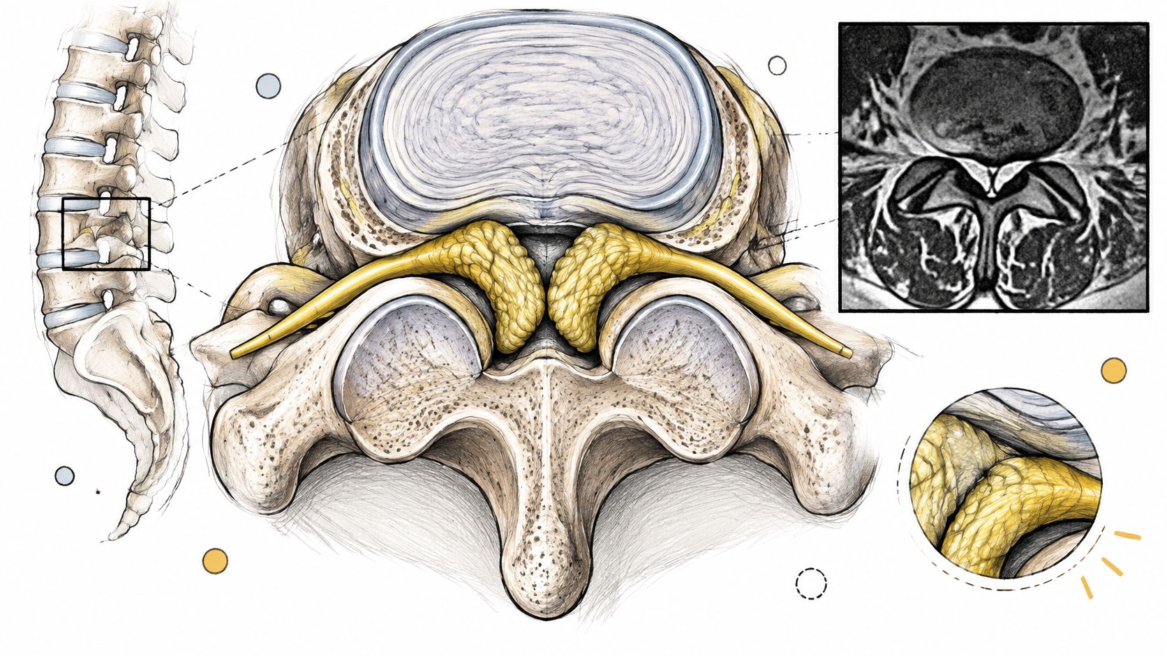
The ligamentum flavum forms part of the posterior boundary of the spinal canal. When it enlarges, it can reduce space around the thecal sac and contribute to narrowing near the lateral recesses, where nerve roots travel. Whether that produces symptoms depends on the location and overall pattern of compression.
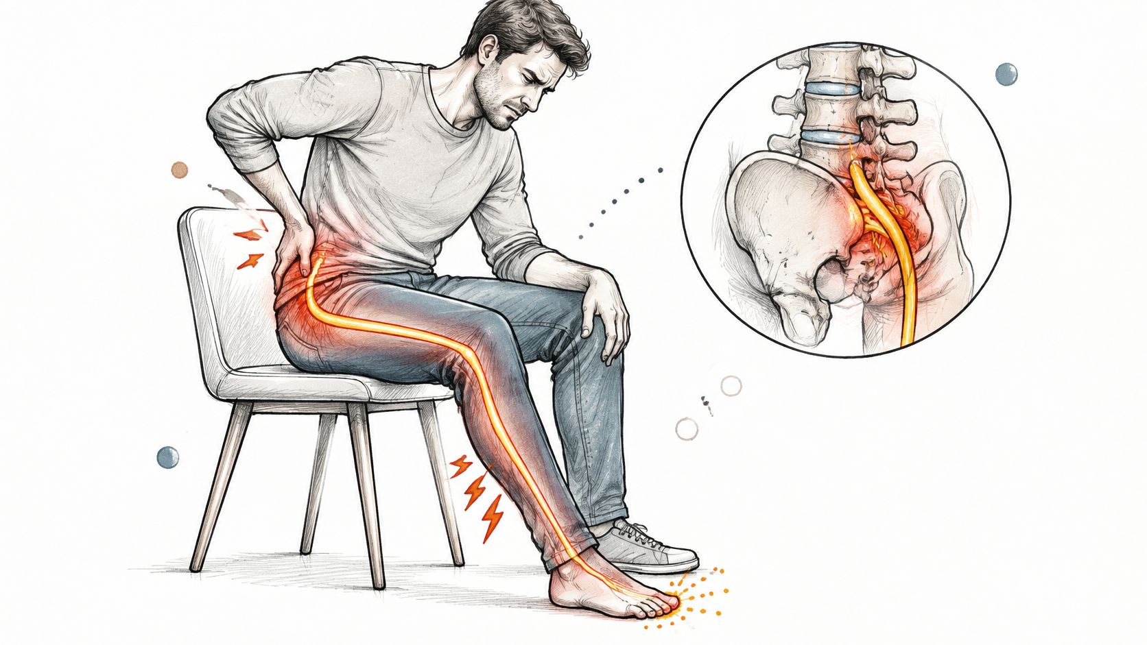
Neurogenic claudication typically involves leg pain or heaviness brought on by walking or standing and relieved by sitting or leaning forward. Radicular pain follows an affected nerve-root distribution and may accompany numbness or weakness. Neither pattern identifies the ligament as the sole cause.
Symptom patterns to match with thickness
| Pattern | Typical clues | What thickness alone cannot show |
|---|---|---|
| Neurogenic claudication | Leg pain or heaviness on walking that eases with sitting or leaning forward | Whether this ligament is the pain generator |
| Radicular pain | Pain along a nerve-root path, with numbness or weakness | Which level is actually compressed |
| Poor symptom correlation | In the older-adult study described above, thickness didn't track walking tests, Pain Disability Index, or visual-analog pain scores | Symptom severity; some asymptomatic participants also had radiologic stenosis and thicker ligaments |
| Red flags | Progressive weakness, gait change, or bladder and bowel symptoms | That it's safe to wait; these require prompt specialist assessment |
Match the symptom to the compressed structure
Say a patient has a report describing bilateral thickening but symptoms affecting only one leg. The next step isn't to assume that both sides need treatment. The reviewer should identify whether a matching nerve root is compressed on the symptomatic side and consider the disc and facet findings at that level.
A symptom diary helps make that comparison concrete:
- Location: Record whether symptoms affect the buttock, thigh, calf, foot, or several regions.
- Trigger: Note whether standing, walking, or a particular posture brings them on.
- Relief: Describe whether sitting or bending forward changes the symptoms.
- Function: Record changes in walking tolerance, balance, or ability to lift the foot.
The clinic's guide to signs of sciatic nerve pain can help patients describe radiating symptoms, but sciatica remains a symptom pattern rather than proof of a particular tissue diagnosis.
Separate nerve-root pain from spinal-cord dysfunction
Lumbar radiculopathy and spinal-cord syndromes shouldn't be grouped together as ordinary back pain. Gait deterioration, hand dysfunction in cervical disease, or progressive weakness may indicate a different level and urgency of neurologic involvement.
New bladder or bowel dysfunction, saddle numbness, or rapidly worsening weakness warrants urgent medical assessment. Don't wait for an elective treatment consultation or a more precise ligament measurement. Clinical deterioration matters even when the report uses a mild-sounding description.
Treatment Pathways From Conservative Care to Decompression
| Pathway | Usually considered when | Main limit |
|---|---|---|
| Conservative care, including physiotherapy, medication, and activity changes | Intermittent symptoms without neurologic decline | Doesn't remove the structural narrowing |
| Targeted injection | Radicular pain that doesn't settle with conservative care | Temporary relief; the duration isn't predictable for an individual |
| Decompression, such as laminectomy or laminotomy | Persistent disabling symptoms or progressive neurologic deficit | Addresses compression but not instability by itself |
| Fusion | Instability or spondylolisthesis, not thickening alone | Adds surgery for instability rather than for thickness |
| Specialist evaluation for thoracic ossification | Clinically significant spinal-cord compression | Conservative care is usually not sufficient once significant ossified compression is established, so specialist evaluation should not be delayed |
Treatment selection follows symptoms, examination, compression, and stability, not a thickness threshold. A ligament measurement cannot show whether the compression is symptomatic or whether instability coexists. The AJNR discussion of ligamentum flavum thickening supports interpreting the ligament alongside facet degeneration and other changes rather than assuming that disc-space narrowing alone explains it.
Choose the intervention for the problem it can address
When symptoms are intermittent and neurologic function is stable, conservative care is usually the starting point. A physiotherapist can tailor exercise, pacing, and movement strategies to the patient's tolerance and examination. For readers arranging rehabilitation at home, Carevo Home Health Care physiotherapy support offers a resource for exploring supervised rehabilitation support.
Medication and targeted injections address symptoms rather than removing the enlarged ligament. An injection may help manage persistent radicular pain, but relief doesn't prove that the ligament was the only contributor, and it shouldn't be described as structural decompression.
Laminectomy or laminotomy creates space around compressed neural structures. A surgeon may consider decompression when symptoms remain disabling despite appropriate nonsurgical care, or when neurologic deficits progress. The decision requires a matching anatomical target at the responsible level.
Fusion answers a separate question: does the segment need stabilization? Instability or spondylolisthesis may influence that decision, but ligamentum flavum thickening alone doesn't establish a need for fusion. A thickness value also cannot settle whether a patient needs surgery; the clinical picture and the level of compression carry that decision.
Recognize when the usual sequence doesn't apply
Thoracic ossification with clinically significant spinal-cord compression isn't equivalent to uncomplicated degenerative lumbar stenosis. Specialist evaluation comes earlier because continued symptom-focused care may leave the cord compressed. The usual ladder from conservative care to decompression assumes a stable neurologic picture, and that assumption fails when the cord is already compressed.
Bring these questions to the surgical discussion:
- Responsible level: Which finding best explains the symptoms and examination?
- Stability: Is there evidence of instability that changes the procedure?
- Decision point: What deterioration or lack of improvement would change the plan?
Decision safeguard: Ask what the proposed treatment changes physically, what it only treats symptomatically, and what it leaves unresolved.
Where Regenerative Stem Cell Therapy Fits
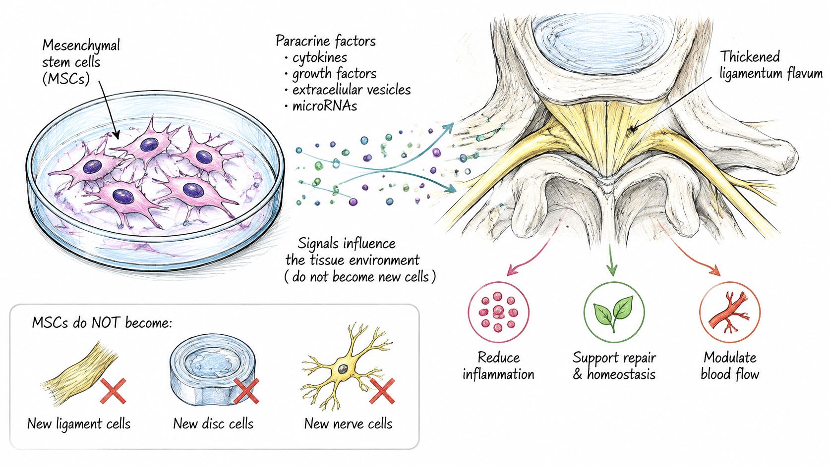
Think of mesenchymal stromal cells, often called mesenchymal stem cells or MSCs, as biological signal senders rather than replacement building blocks. In the therapeutic mechanism discussed here, they don't turn into new ligament, disc, or nerve cells. Their proposed main action is the paracrine effect, releasing specialized signals that influence inflammation, repair responses, vascular activity, and nearby cells.
The diagram illustrates this signaling model.
Positive findings need the correct clinical context
A review of clinical MSC evidence in spinal-cord injury describes a randomized trial in which 34 patients received a single intramedullary dose of 4 × 10⁷ allogeneic umbilical-cord MSCs, with reported improvements in motor activity, muscle tone, Barthel Index, and urodynamic measures at six months. The same review describes 10 patients receiving four intrathecal doses of 30 × 10⁶ autologous bone-marrow MSCs, with reported sensory, motor, and urodynamic improvements and reduced neuropathic pain at 12 months.
These are encouraging findings in spinal-cord injury, not trials of ligamentum flavum thickening. They support investigation of signaling-based treatment but don't demonstrate that MSCs shrink a fibrotic ligament, reverse ossification, or relieve mechanical lumbar stenosis. A proposed mechanism is not condition-specific proof.
This distinction is especially important when leg pain has several possible sources. Regenerative treatment aimed at an associated joint or soft-tissue problem shouldn't be presented as equivalent to removing tissue that compresses a nerve.
The accompanying video provides additional clinic context.

Place the option beside the diagnosis
At Dream Body Clinic, our lower-back treatment information covers herniated discs, degenerative disc disease, facet-joint arthritis, and sciatica. Our injection site depends on whether the problem is a ligament or tendon injury or arthritis; patients can review our stem cell therapy for sciatica information when preparing for case review.
These treatments are delivered in Mexico within the COFEPRIS-regulated framework. That operating context doesn't by itself establish authorization or proven effectiveness for every indication, and the sources cited here don't establish clinic-specific approval for treating ligamentum flavum thickening.
The appropriate role is an informed, diagnosis-specific discussion, not an assumption that any thickened ligament is suitable for regenerative care. Progressive neurologic compression still requires timely spine-specialist assessment.
Confirm Your Imaging Before Booking a Treatment
An isolated MRI report can't predict your individual course. Age-related degeneration may coexist with stable function, while a change in strength or walking can make reassessment important even without a new thickness measurement. Prognosis depends on the clinical trajectory and the structures involved.
Prepare a decision-ready imaging record
Obtain the complete MRI. Bring the images and report, with level-by-level findings and both sides where relevant. A summary phrase alone may omit the anatomical detail needed for treatment selection.
Clarify any recommendation for CT. If mineralization or an unusual lesion is suspected, resolve that question before choosing an intervention.
Write down the symptom pattern. Include walking and standing triggers, positional relief, numbness, weakness, and any bladder or bowel change. Flag deterioration rather than burying it in a general pain history.
Ask for a level-specific explanation. Establish which finding matches the examination and whether instability changes the options.
Compare treatments against that target. Ask what improvement is realistic, how it will be measured, and what findings would require another pathway.
For broader imaging context, Longevity Clinics on full body MRI discusses screening, but screening isn't a substitute for targeted spine imaging and neurologic assessment.
Verify the treatment and facility separately
The AABB summary of Mexico's cellular-therapy framework identifies COFEPRIS as the responsible health regulator and explains establishment licensing requirements for handling tissues and cells. Regulatory oversight isn't blanket approval of every intervention for every condition; applicable facility and treatment documentation still matter.
Claims about repairing facet cartilage, ligaments, tendons, or muscles shouldn't be interpreted as evidence that a treatment reverses ligamentum flavum stenosis. Bring your imaging and authorization questions to a complimentary case review, and make the decision from the diagnosis rather than the measurement.
Dream Body Clinic offers regenerative treatment consultations for herniated discs, ligament or tendon injuries, and facet-joint arthritis, conditions that may coexist with ligamentum flavum thickening. Visit Dream Body Clinic to request a complimentary case review with your MRI images, report, and symptom history, so the discussion starts with whether your findings match an appropriate treatment target.