You've opened your MRI report late at night, searched every unfamiliar term, and now you have two labels that seem to describe the same problem. One line says degenerative disc disease, another says spondylosis, and neither explains why sitting hurts, why your leg tingles, or whether an injection could help. That confusion isn't a failure on your part. Those words describe overlapping anatomy, not a single pain source, and the difference between them changes what treatment can plausibly do.
Table of Contents
- Why the Words on Your MRI Report Change Your Options
- What Spondylosis Means and How It Goes Beyond the Disc
- Comparing DDD and Spondylosis Side by Side
- How Doctors Tell the Two Apart on Imaging
- Conventional Care From Conservative Treatment to Surgery
- How MSC Therapy Is Thought to Work in the Spine
- What the Evidence Actually Says About Outcomes
- Making the Right Choice for Your Situation
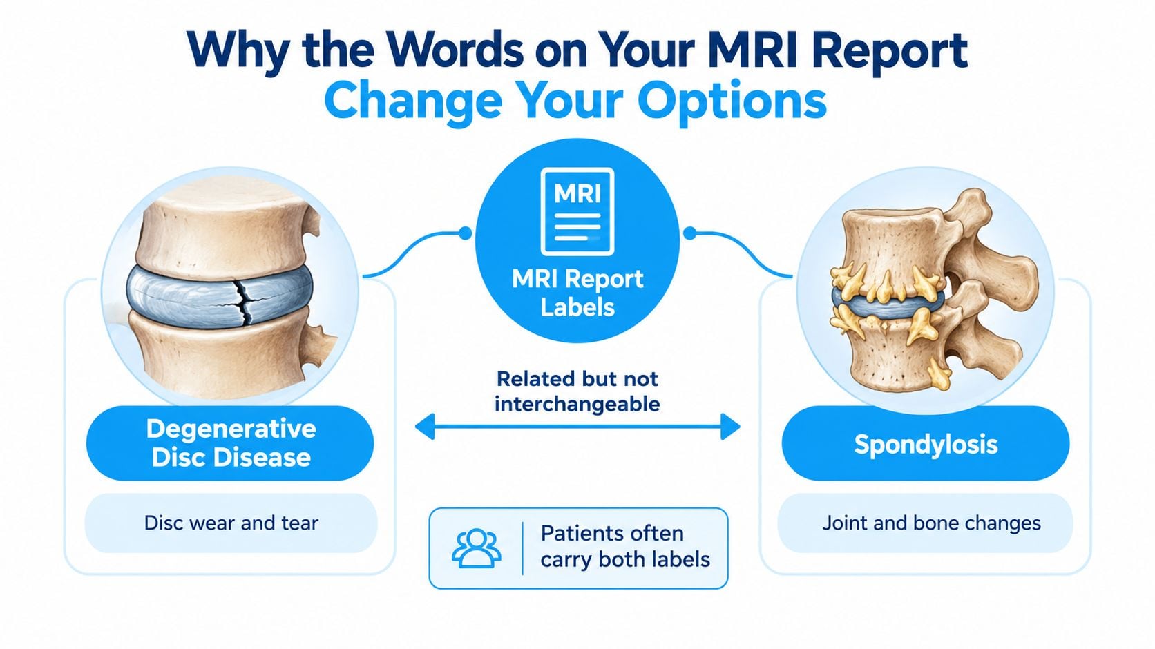
Why the Words on Your MRI Report Change Your Options
The individuals I review cases with aren't asking for a lecture on anatomy. They want to know three practical things. Is this finding causing my pain, can it be treated without surgery, and what would improvement look like? Degenerative disc disease and spondylosis point to different answers, even when both appear on the same report.
What degenerative disc disease describes
Degenerative disc disease, often shortened to DDD, primarily describes structural failure inside an intervertebral disc. The disc loses water, which radiologists call desiccation and see as reduced T2 signal on MRI. Height drops, the outer annulus can fissure, and the vertebral endplates next to the disc can remodel.
A healthy disc spreads load across the segment. A dehydrated, narrowed disc does not. Load shifts backward toward the facet joints and forward in uneven patterns, and the openings where nerves exit can become smaller because the segment has collapsed. That mechanics story matters, but it remains an anatomy description. It does not, by itself, prove the disc is generating your pain.
Practical rule: Treat DDD on a report as a finding to correlate, not as a verdict. The useful question is whether your pain pattern, examination, and function match that level.
Age explains a large part of the caution. In a systematic review of 33 studies involving 3,110 asymptomatic people, disc degeneration increased from 37% at age 20 to 96% at age 80, and disc bulging increased from 30% to 84% across the same ages. People with no symptoms carried these findings often, and the frequency rose steadily with age.
That doesn't mean degeneration is irrelevant. It means a clinician can't responsibly say, "Your disc looks worn, therefore that's your pain," and then sell a procedure on that logic alone. Axial pain in the middle of the low back, pain that travels in a clear nerve pattern, facet mediated pain, muscle pain, and non spinal causes can overlap. They call for different next steps.
Why this matters beyond one report
The scale of back pain is one reason sloppy labeling causes real harm. In the Global Burden of Disease Study 2021 estimates for global low back pain00098-X/fulltext), there were 619 million prevalent cases in 2020, with a 95% uncertainty interval of 554 to 694 million, and low back pain accounted for 7.7% of all years lived with disability, making it the world's leading contributor to disability. Not every one of those cases is caused by DDD or spondylosis, and that distinction is the point.
When labels are used loosely, patients get pushed toward loose decisions. They chase MRI severity instead of functional goals, repeat injections that were never well targeted, or assume a regenerative treatment will rebuild every structure named on the report. A better starting vocabulary prevents that.
Use these questions when you read any report:
- Which structure is named: Is the finding mainly in the disc, or does it also involve bone spurs, facet joints, ligaments, and narrowing?
- Which level is involved: Does that spinal level match where you feel pain, numbness, or weakness?
- Is a nerve or the cord compressed: Wear alone is different from compression with matching neurological signs.
- What's limited in daily life: Walking distance, sitting tolerance, sleep, grip, and work tasks often matter more than a single image grade.
DDD tells you the disc has changed. Spondylosis, covered next in anatomical terms, tells you the degenerative process has spread beyond the disc. Once you separate those ideas, candidacy for physical therapy, injections, surgery, or mesenchymal stromal cell therapy becomes much clearer.
What Spondylosis Means and How It Goes Beyond the Disc
Spondylosis is the broader term. Build the picture outward from the disc. First the disc dries and loses height. Then the segment moves and loads differently, and surrounding structures respond over time.
The structures added by spondylosis
- Osteophytes: Bony spurs form at vertebral margins as the body tries to stabilize a segment that's loading unevenly.
- Facet joint arthropathy: The paired joints at the back of the segment develop cartilage wear, swelling, and bony enlargement, often called facet hypertrophy.
- Ligamentum flavum thickening: A ligament along the back of the canal thickens and can buckle inward, taking up space.
- Canal or foraminal narrowing: The central canal, lateral recess, or nerve exit openings become smaller, which clinicians describe as stenosis at specific levels.
Sleep position won't reverse those changes, but neck support can reduce overnight strain while you're sorting out the larger plan, and it's reasonable to find your neck pain relief pillow as part of basic symptom management. For readers whose neck pain localizes to the back of a segment, understanding the anatomy and role of the cervical facet joint helps explain why facet arthropathy and disc desiccation aren't the same target.
Spondylosis becomes clinically important when those changes encroach on neural tissue. A foraminal osteophyte complex can compress one exiting nerve root and produce arm or leg symptoms in that root's pattern. Central narrowing in the neck can threaten the spinal cord. Without encroachment and matching signs, multilevel spondylosis may describe an aging spine that isn't the main pain generator.
Comparing DDD and Spondylosis Side by Side
A side by side view prevents the most common mistake, treating both labels as interchangeable reasons for the same procedure. The contrast is about anatomy, likely symptom pattern, and what makes each finding actionable.
DDD vs. Spondylosis at a Glance
| Criterion | Degenerative Disc Disease | Spondylosis |
|---|---|---|
| Primary anatomical target | Intervertebral disc, including nucleus, annulus, and adjacent endplates | Disc plus vertebrae, facet joints, ligaments, and bony margins |
| Typical pain pattern | Axial neck or low back pain when symptomatic, sometimes with disc related referred pain | Axial pain, facet mediated pain, radicular pain, or myelopathy depending on narrowing |
| Imaging hallmarks | Reduced T2 signal, disc height loss, annular fissuring, bulge or protrusion, endplate changes | Osteophytes, facet arthropathy and hypertrophy, ligament thickening, foraminal or central stenosis |
| When it becomes important | When symptoms, examination, and level line up and function is limited | When bony or soft tissue narrowing compresses a root or cord, or facet disease matches the pain |
| Usual first pathway | Conservative care for uncomplicated axial pain, with targeted escalation if concordance is strong | Conservative care without neurological compromise, prompt specialist review when compression signs appear |
Imaging alone can't settle which column explains your symptoms. In summarized MRI findings among asymptomatic adults, reduced disc signal intensity appeared in approximately 20% to 83%, disc bulges in 10% to 81%, disc protrusions in 3% to 63%, disc narrowing in 3% to 56%, and annular tears in 6% to 56%. Those wide ranges show why a structurally abnormal disc can be clinically silent.
In practice, isolated disc degeneration with axial pain and a normal neurological examination is a different decision problem from multilevel spondylosis with foraminal stenosis and matching leg weakness. If you're reviewing conventional disc pathways before considering regenerative care, Pain Management of Williamsport disc care outlines how degenerated or herniated discs are commonly framed in pain practice. The table gives you the categories. Clinical correlation decides the pathway.
How Doctors Tell the Two Apart on Imaging
Good imaging interpretation starts before the scan. MRI is sensitive, which is useful when there's a specific question and misleading when there's none. Uncomplicated spine pain without warning signs doesn't automatically need early MRI, because incidental degeneration can create anxiety and pull care toward procedures that don't improve function.
The five step view below shows how a careful workup moves from restraint to correlation.
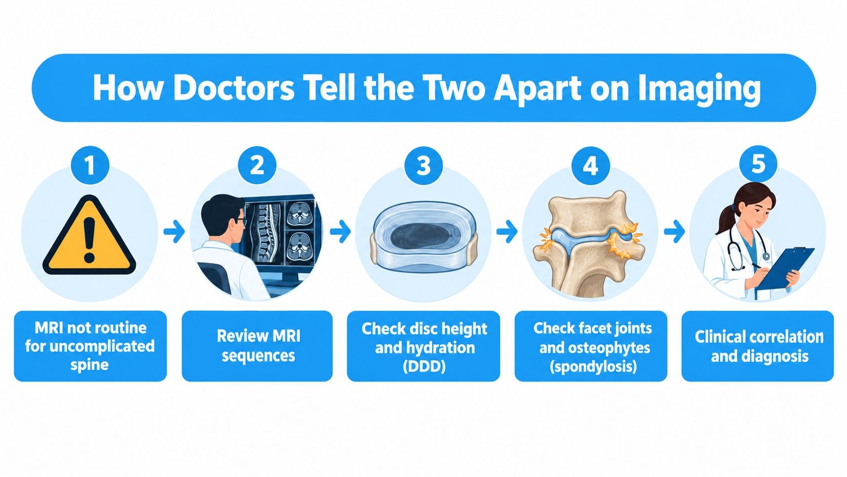
When MRI becomes appropriate
Persistent radiculopathy, progressive weakness, saddle anesthesia, bowel or bladder dysfunction, and suspected myelopathy change the urgency. In the neck, gait disturbance, hand clumsiness, hyperreflexia, and pathologic reflexes are long tract signs that shouldn't be managed as routine wear and tear. Those findings require prompt specialist assessment.
On the images, T2 signal loss inside a disc points to desiccation. T2 hyperintensity inside the spinal cord is a different signal in a different tissue, and it shouldn't be brushed off as ordinary spondylosis. In cervical spine guidance on reporting degenerative imaging by level and mechanism, severity should be specified as disc protrusion, osteophyte complex, facet hypertrophy, foraminal stenosis, central stenosis, or cord involvement, because cord hyperintensity may reflect edema, inflammation, ischemia, myelomalacia, or gliosis.
Ask for that level by level language. Then pair it with dermatomal pain, sensory change, reflex asymmetry, myotomal strength, walking tolerance, and a disability score such as the Oswestry Disability Index. A report that can't be matched to examination findings hasn't yet identified a treatment target.
Conventional Care From Conservative Treatment to Surgery
Regenerative options make more sense when you see the conventional ladder they sit beside. The early steps aren't glamorous, but they're where many uncomplicated cases improve, and they also create the baseline that tells you whether escalation is justified.
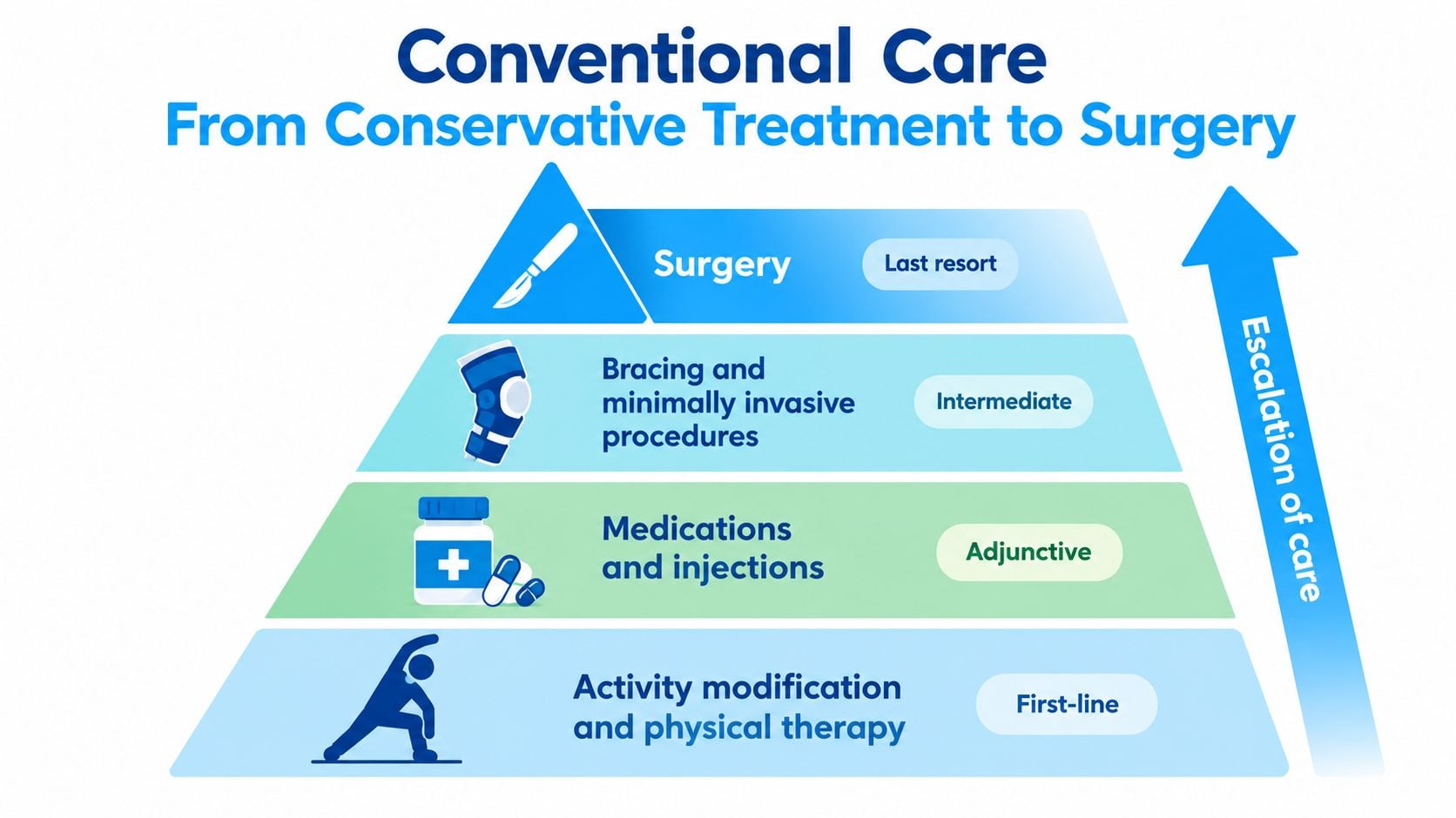
The usual escalation path
- Conservative care first: Activity modification, structured physical therapy, anti inflammatory medication when appropriate, sleep improvement, and weight management where relevant.
- Targeted injections: Epidural steroid injections for selected radicular pain, or facet joint injections and medial branch procedures when facet mediated pain is suspected.
- Radiofrequency ablation: Considered for facet pain that responds temporarily to diagnostic blocks, not as a generic answer to a worn disc.
- Surgery for clear indications: Decompression or fusion enters the discussion when concordant compression, progressive deficit, myelopathy, or failed conservative care supports it.
That sequence reflects guidance discouraging routine imaging for uncomplicated low back pain, which warns that incidental abnormalities can trigger anxiety and unnecessary procedures, and notes that for suspected acute disc herniation without red flags, conservative care is generally tried before MRI with physical therapy first. For suspected herniation, a common example is roughly six weeks of therapy before imaging, but your own clinician should set the timing.
Trade offs stay visible here. Repeated epidural steroids without a clear radicular target, and fusion for nonspecific axial degeneration, sit in a grayer area than decompression for clear myelopathy. Readers weighing leg dominant symptoms can review how regenerative treatment is framed for sciatica related pain, but surgery shouldn't be deferred when progressive neurological signs demand it.
How MSC Therapy Is Thought to Work in the Spine
Marketing language most often outruns biology. Mesenchymal stromal cells, or MSCs, aren't credibly presented as injected building blocks that move in and become a new disc. The stem cells don't differentiate into other cells as the main explanation for benefit, and they don't need to engraft permanently to matter in the current model.
The diagram below places the proposed signaling role at the center of the explanation.
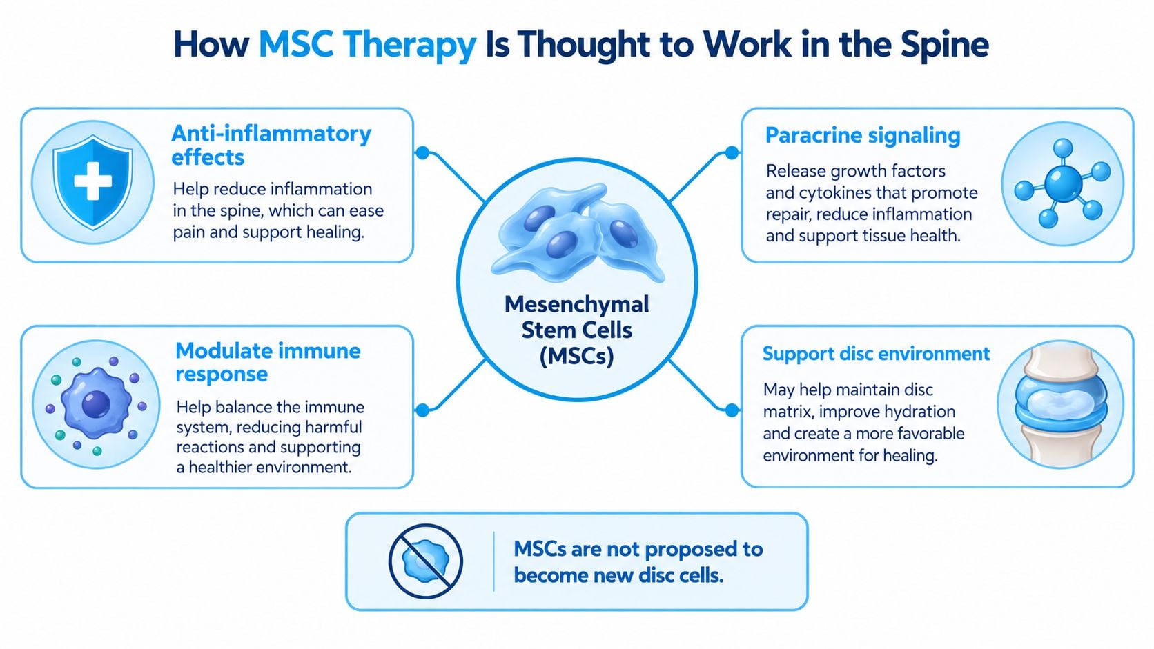
Paracrine signaling explained plainly
In research describing the paracrine model of MSC action in degenerative discs, MSCs are proposed to work mainly through paracrine signaling, releasing soluble factors and extracellular vesicles that communicate with resident disc cells. Reported effects include stimulation of extracellular matrix production, reduced inflammatory signaling, and protection against cell death, with the therapeutic effect thought to depend more on released signals than on engraftment or differentiation into new disc cells.
The cells send specialized signals that guide the local healing process. Your remaining disc cells, not a newly manufactured disc, do most of the responding. That is a more modest claim than regeneration in the everyday sense, and modesty matters when you are deciding what to pay for and what outcome to expect.
Specific molecules named in recent work on MSC signaling in disc degeneration include interleukin 10 and transforming growth factor beta, alongside trophic factors such as insulin like growth factor 1 and growth and differentiation factor 5. Those signals have been associated with suppression of matrix degrading enzymes such as MMPs and ADAMTS, inhibition of apoptosis, and support for residual disc cells resuming matrix production.
The short video below gives a visual introduction before you compare that mechanism with injections or surgery.

Compared with steroids, which mainly suppress inflammation for symptom relief, MSC therapy aims to alter the disc microenvironment. Compared with surgery, it doesn't remove an osteophyte, enlarge a canal, or stabilize a segment mechanically. If you want the broader delivery picture, read a plain language explanation of how stem cell injections are intended to work and then judge any spine protocol against that mechanism.
MSCs are best understood as signaling cells. They aren't a spare parts service for bone spurs, thickened ligaments, or a compressed spinal cord.
That distinction drives candidacy. A signaling based therapy fits a biologically active, painful disc environment more plausibly than advanced spondylosis where the dominant problem is fixed bony narrowing.
What the Evidence Actually Says About Outcomes
Mechanism creates plausibility. Trials decide confidence, and the current trial record for intradiscal MSC therapy is early and mixed. That isn't a reason to dismiss the field. It's a reason to separate safety signals, structural measurements, and the outcomes patients actually feel.
In the 2025 trial and review evidence summarized for intradiscal MSC treatment, a phase IIB randomized controlled trial found intradiscal bone marrow MSCs were safe and improved disc height measurements, yet they did not outperform sham treatment for pain or disability at six months. A 2025 review identified only 10 studies published from 2011 to 2025, totaling 736 participants, and emphasized small samples and moderate risk of bias.
How to read that result without hype
Improved disc height without better pain scores tells you structure and symptoms don't move in lockstep, a theme you've already seen in asymptomatic MRI findings. A disc can measure slightly better while the person doesn't walk farther, sit longer, or need less medication. Conversely, a person can report better function without a dramatic MRI reversal, because inflammation and pain processing aren't captured by height alone.
For clinic decisions, define success before treatment:
- Pain: Track a numerical pain rating in the same activities that currently provoke symptoms.
- Function: Record walking tolerance, sitting time, lifting, sleep interruption, and a validated disability score.
- Neurology: Document strength, sensation, and reflexes when radicular symptoms exist.
- Medication and work: Note whether daily functioning improves, not only whether an image looks different.
The most plausible near term benefits in carefully selected disc cases are reduced inflammatory signaling, slower matrix breakdown, and modest symptom improvement. Reliable reversal of multilevel spondylosis, facet arthritis, and stenosis hasn't been established. If a clinic guarantees regeneration of every structure on your report, that promise has moved beyond the evidence.
Making the Right Choice for Your Situation
Start with presentation, not with a product. Four patterns cover most consultations for degenerative disc disease and spondylosis, and each leads somewhere different.
Match the pathway to the presentation
Isolated axial pain without neurological signs is the clearest case for continued conservative care first. If pain persists, the pain generator is reasonably concordant with a disc level, and there's no significant compression, an MSC based protocol can be discussed with the realistic goal of paracrine support rather than MRI reversal. That discussion should include dose, delivery route, product source, follow up cadence, and how response will be measured.
Radiculopathy with concordant imaging requires more restraint. Dermatomal pain, sensory change, reflex asymmetry, or weakness that matches a compressed root may respond to conservative care or a targeted epidural approach, but progressive deficit needs surgical opinion. A signaling therapy doesn't physically decompress a root pinched by an osteophyte complex, even if inflammation around that root contributes to pain.
Multilevel spondylosis with canal or foraminal stenosis is where expectations need the tightest control. Bone spurs, facet hypertrophy, and ligament thickening create a space problem. MSC signals don't shave bone or reopen a foramen. Some patients still pursue regenerative care for adjacent disc or inflammatory components, but they should understand that the stenotic component may continue to limit walking regardless.
Suspected myelopathy isn't a watch and inject category. Gait disturbance, hand clumsiness, hyperreflexia, spreading numbness, saddle anesthesia, or bowel and bladder dysfunction require urgent specialist evaluation. Travel for elective regenerative care shouldn't delay that assessment.
Questions to ask any clinic
- What's my presumed pain generator: Ask which finding explains your specific symptoms and what examination evidence supports it.
- What won't this treatment do: A credible answer names limits around bone spurs, fixed stenosis, instability, and cord compression.
- What's the exact protocol: Request the MSC source, stated dose, delivery route, imaging guidance, and whether platelet rich plasma or other adjuncts are included.
- How will you judge response: Look for baseline and follow up pain, disability, walking, strength, and medication measures, not testimonials alone.
- What are the risks and regulatory details: Ask how product quality is controlled and what happens if symptoms worsen after you return home.
Safety questions aren't pessimism. In the FDA consumer alert on regenerative medicine products, products marketed without appropriate approval may be associated with serious risks, and effectiveness, product quality, administration route, and safety depend on the specific protocol and regulatory status. Reported concerns in that broader warning include infection, inflammatory or immune reactions, neurological events, and unintended tissue growth. A clinic should discuss those issues directly rather than calling treatment risk free.
International patients often compare options across regulatory systems. Treatments delivered under Mexico's COFEPRIS framework can include protocols not commonly available in the United States or Canada, which is context, not proof of benefit. A published protocol with transparent dosing, screening, and remote follow up is a better quality signal than broad claims. Dream Body Clinic, based in Bucerias, Nayarit, offers imaging screened lower back MSC protocols for conditions including degenerative disc disease and facet joint arthritis, with complimentary case review, in clinic treatment typically lasting one to three hours, and remote check ins at approximately three, six, and twelve months. Treat that structure as one model to compare against other clinics using the checklist above.
Daily habits still matter while you decide. Activity pacing, sleep, weight management, and physical therapy adherence influence symptoms regardless of injections, and readers who prefer non drug adjuncts can explore science-backed natural pain relief as supportive reading rather than a substitute for diagnosis. Those measures won't correct concordant cord compression, and they shouldn't be used to postpone urgent care.
Quick answers for traveling patients
Does an MRI finding alone make me a candidate? No. Candidacy requires concordance between the finding, your symptoms, neurological examination, and functional limits. A silent degenerative disc doesn't need a procedure because it looks dramatic.
What outcome is realistic if I'm selected? The reasonable goal is improved pain and function over months, possibly with a healthier disc environment, not guaranteed restoration of a youthful spine. If your main limitation comes from fixed spondylotic narrowing, set expectations accordingly.
How is follow up usually structured after travel? Published international protocols commonly use remote reviews around three, six, and twelve months, with repeat imaging when clinically indicated. Confirm who reviews deterioration, who coordinates care with your home clinician, and what thresholds trigger urgent local evaluation.
The right choice is rarely the clinic with the boldest promise. It's the pathway that names your pain generator accurately, respects red flags, measures function, and explains mechanism without turning signaling into a guarantee.
If your MRI mentions degenerative disc disease, spondylosis, facet arthritis, or stenosis and you want a candidacy review grounded in concordance rather than imaging alone, visit Dream Body Clinic to request a complimentary case review. Share your MRI report, neurological symptoms, and functional goals so the team can tell you whether a lower back or neck MSC protocol fits your presentation, what it can't address, and how follow up would be structured after treatment in Bucerias.