
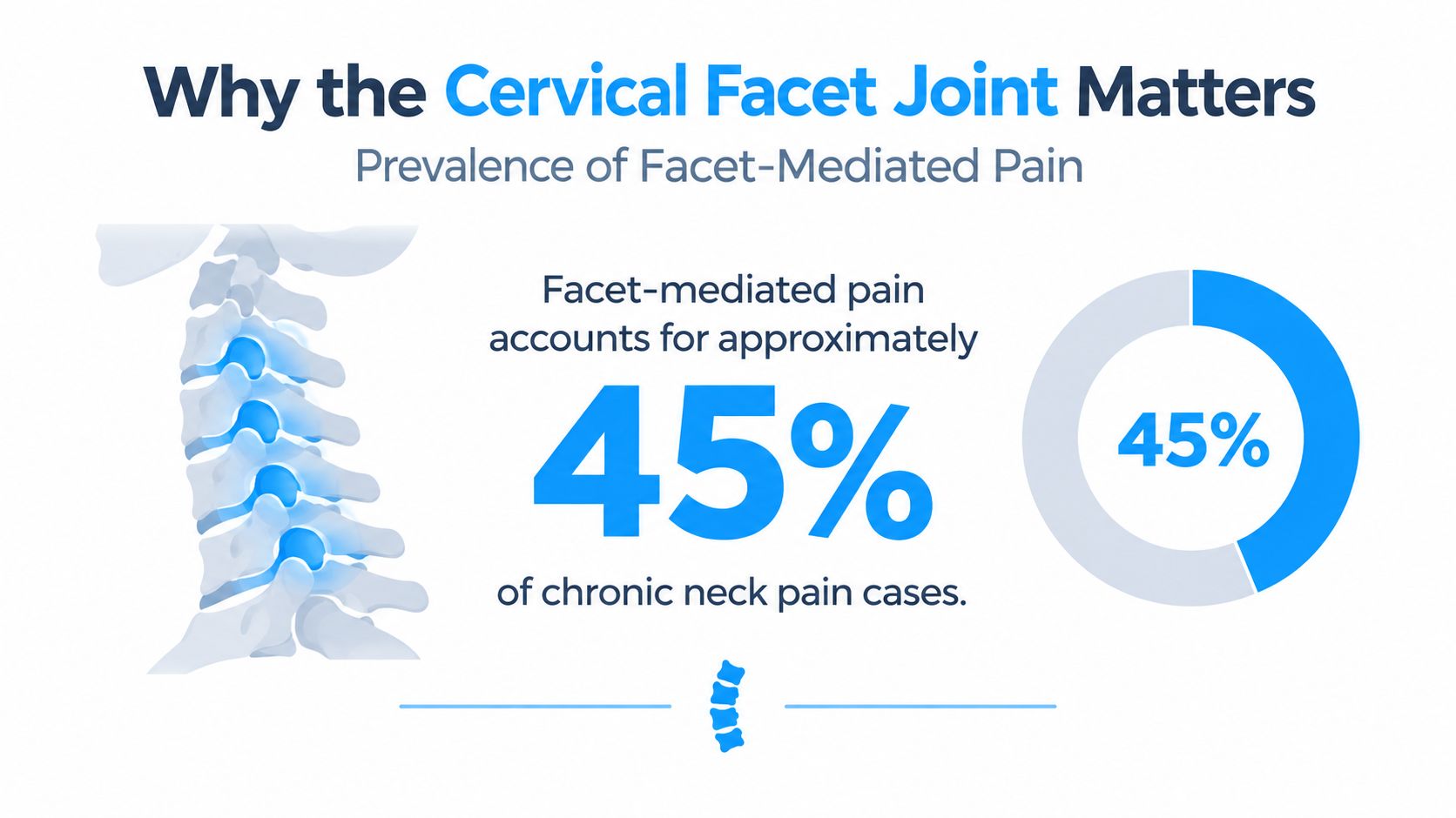
Facet-mediated pain accounts for 26% to 70% of chronic neck pain cases, and in whiplash-associated neck pain it's still a major culprit, not a rare exception (Pain Medicine consensus guidelines). That single fact changes how you should think about a stiff, painful neck, because the problem often lives in the cervical facet joints, not in the muscles alone. These are the small paired joints at the back of the neck that guide motion, share load, and can become painful long before a scan gives you a simple answer.
Table of Contents
- Why the Cervical Facet Joint Matters
- Anatomy and Function of Cervical Facet Joints
- What Is Cervical Facet Arthropathy
- Symptoms and Clinical Signs to Recognize
- How Cervical Facet Pain Is Diagnosed
- Conventional Treatment Options From Conservative to Ablative
- Regenerative Injections and the Paracrine Mechanism
- Comparing Regenerative Therapy to Conventional Options
- What a Regenerative Treatment Journey Looks Like
- Quick Reference for Patients and Clinicians
- Frequently Asked Questions About Cervical Facet Joint Pain
Why the Cervical Facet Joint Matters
A cervical facet joint is one of the paired joints that connects one vertebra to the next. Think of it as a small bony knuckle lined with cartilage and wrapped in a capsule, sitting behind the disc and separate from the uncovertebral joint. Its job is simple but critical, it keeps the neck stable while still letting you turn, nod, and tilt.
That matters because the neck is used constantly. Every time you look over your shoulder, check a blind spot, lift your head from a pillow, or hold your phone too low, these joints share the mechanical load. When they become irritated, even ordinary movement can start to feel sharp, tight, or restricted.
Why this joint gets overlooked
Many people hear “neck pain” and assume the disc is the problem. Others think the muscles must be the only issue because the pain feels tight or stiff. In reality, the cervical facet joint is its own pain source, and it often sits at the center of chronic axial neck pain, whiplash symptoms, and age-related stiffness.
Practical rule: if your neck pain is worse with motion than with rest, the facet joints deserve attention.
That's why this topic matters before you ever get to injections or regenerative care. If you don't know the anatomy, it's easy to misread the symptoms and chase the wrong structure.
Anatomy and Function of Cervical Facet Joints
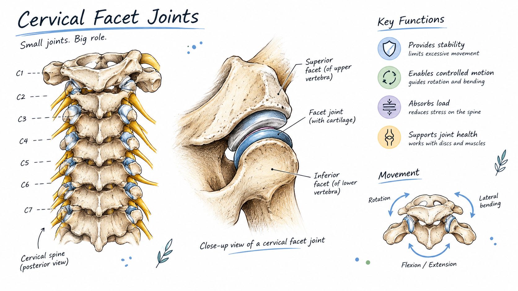
The cervical facet joints are paired synovial diarthrodial joints in the back of the neck, running from C2–3 to C7–T1. Each joint is made by the superior and inferior articular processes of neighboring vertebrae, with cartilage, synovial fluid, and a capsule that lets the joint surfaces glide instead of grind (Oxford textbook chapter).
How the neck balances motion and stability
Their shape changes level by level. The upper cervical facets sit more horizontally, while the lower levels become more oblique and gradually shift toward a sagittal alignment. That geometry helps the neck rotate, bend, and still remain stable. The joints are innervated by the medial branches of the dorsal rami, and one facet joint usually receives input from two nearby levels. That is why clinicians often assess two nerves when they test one joint.
A useful mental picture is a row of small knuckles, each with its own capsule and lining. When one knuckle becomes irritated, the movement pattern of the whole neck can feel off.
Why imaging needs context
Plain X-rays can show alignment, but they do not show every detail well because these joints sit at an angle. CT and MRI give a better multiplanar view, so doctors use them for anatomical planning and to look for degeneration or other structural change. Even so, imaging cannot prove that a specific facet joint is the pain source, because the neck can show wear without matching symptoms.
That limitation matters in real practice. Pain from a cervical facet joint is confirmed by targeted diagnostic blocks, and even those blocks can produce false-positive results, which is why treatment decisions need both anatomy and careful clinical testing. A scan is useful information, not a verdict.
What Is Cervical Facet Arthropathy
Cervical facet arthropathy is the degenerative change of the facet joint. It usually starts with irritation of the joint lining and capsule, then can progress to cartilage loss, bony thickening, joint irregularity, and, in advanced cases, fusion. In plain language, the joint becomes worn, stiff, and less smooth.
That wear is common enough that scans alone can be misleading. The neck may show degeneration even when symptoms do not match, so the diagnosis still has to be anchored in the pain pattern, exam findings, and response to targeted blocks. False-positive diagnostic blocks can also steer treatment in the wrong direction, which is why a scan should be treated as context, not proof.
What the scan can show, and what it can't
CT and MRI can show degeneration, but they cannot confirm that one specific facet joint is the pain source. That distinction matters because structural change and pain do not always track together. A joint can look worn without being the main reason a patient hurts.
That is why clinicians still rely on diagnostic blocks, while also recognizing their limits. A block can calm pain from the intended joint, but it can also give a false-positive result, especially when nearby structures contribute to symptoms. Real-world decisions depend on both anatomy and careful testing, not imaging alone.
For people who want a broader patient-friendly overview of joint degeneration and symptom control, natural osteoarthritis remedies can help frame conservative care alongside medical evaluation. For patients considering regenerative care for spine degeneration, the cervical facet question often overlaps with broader stem cell therapy for osteoarthritis.
Degenerative changes in cervical facet arthropathy
| Stage | Pathology | Typical Imaging Finding |
|---|---|---|
| Early | Synovitis and capsular stretch | Subtle joint-space irritation or no obvious change |
| Moderate | Cartilage loss and bony remodeling | Joint-space narrowing, osteophytes |
| Advanced | Marked degeneration and stiffness | Irregularity, sclerosis, fusion |
Facet arthropathy is not the same as disc degeneration, and it is not an autoimmune arthritis diagnosis by default. It is a mechanical degenerative pattern, and that frame keeps treatment choices grounded.
Symptoms and Clinical Signs to Recognize
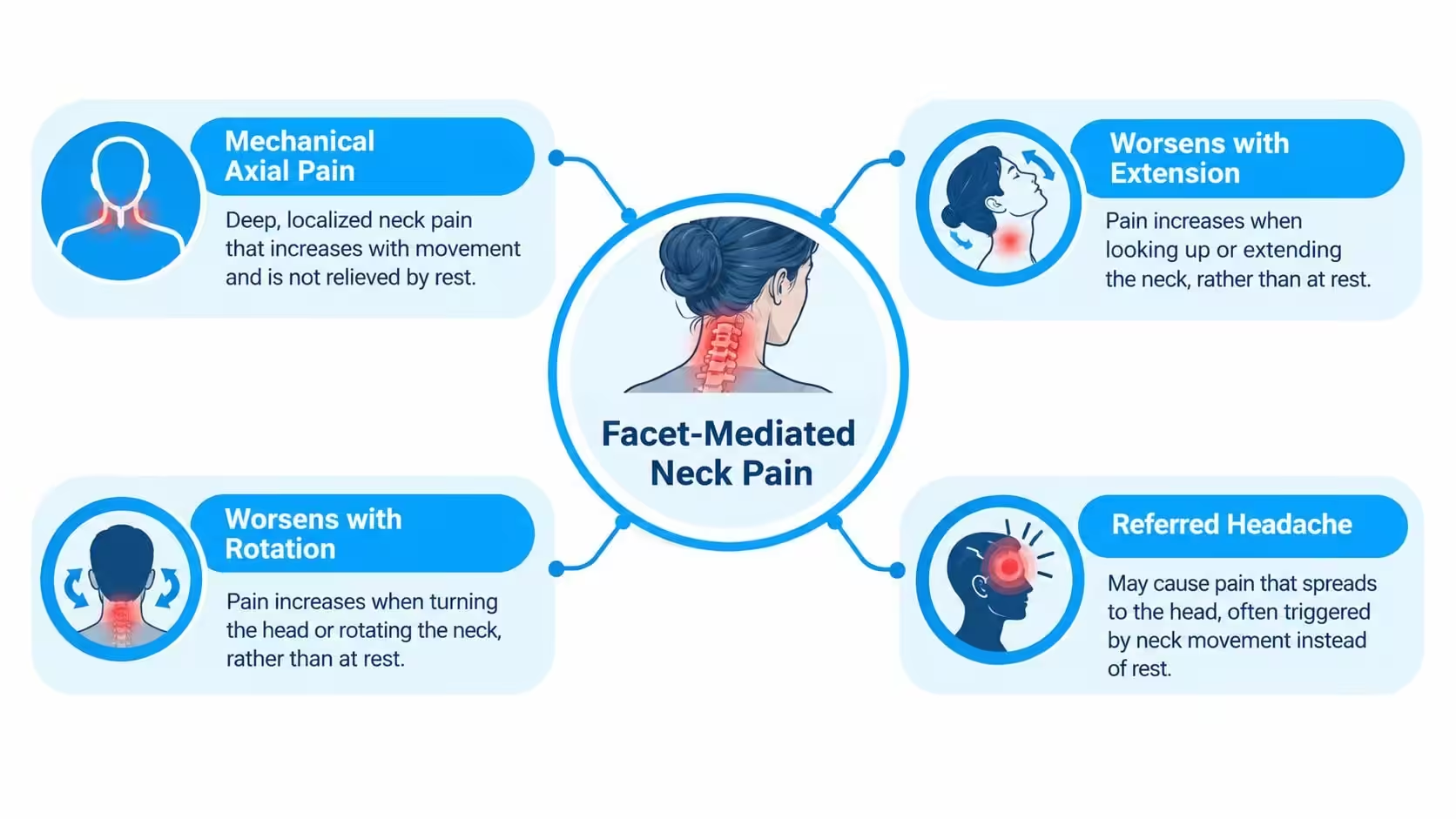
Facet-mediated pain usually feels like mechanical axial neck pain. The pain often worsens with extension and rotation, because those movements load the posterior joints more than flexion does. People often describe a deep ache, stiffness, or a sharp catch when turning the head.
Where the pain travels
Upper cervical joints can refer pain into the occipital region, the upper trapezius, the shoulders, and the periscapular area. That's why C2-3 irritation can look like occipital neuralgia, even when the nerve itself isn't the main problem. Lower cervical levels more often send discomfort toward the shoulder girdle and upper back.
Whiplash changes the picture too. The joint capsule and small stabilizing ligaments can be injured in a way that doesn't always show up clearly on standard imaging, yet the pain can persist and flare with movement.
What clinicians look for
- Extension-rotation loading, because it compresses the joint and can reproduce concordant pain.
- Ipsilateral rotation, which may trigger the same ache a patient feels during daily activities.
- Localized tenderness, especially when the pain map matches a facet referral pattern.
- Absence of nerve-root signs, because radicular arm pain points clinicians toward a different source.
If the pain shoots down the arm, causes numbness or weakness, or comes with myelopathic or systemic symptoms, facet pain stops being the leading explanation.
Symptom pattern raises suspicion, but it doesn't confirm the joint as the source. That uncertainty is exactly why the diagnostic process has to be more precise.
How Cervical Facet Pain Is Diagnosed
The first diagnostic mistake is assuming a scan can do the whole job. X-ray, CT, and MRI can show osteophytes, cartilage loss, capsular thickening, and other structural changes, but those changes don't reliably identify the painful joint by themselves. That's why imaging helps with anatomy and planning, not with certainty about pain generation.
Why medial branch blocks matter
Controlled medial branch blocks are widely treated as the reference standard for diagnosis. The reason is anatomical, each cervical facet joint is supplied by two medial branches from adjacent levels, so blocking the right nerves can temporarily interrupt pain from that joint. If pain falls in a clear, controlled way, the joint becomes a much more credible culprit.
Single uncontrolled blocks can mislead clinicians. Recent synthesis reports reported prevalence ranges from 36% to 67% at ≥ 80% pain relief, with estimated false-positive rates from 26% to 63%, which is why dual blocks are often used to reduce misclassification (AAH repository review).
What the test really means
A positive block doesn't mean the joint is “the only problem.” It means the probability of facet-mediated pain is high enough to justify the next step. That's a more honest way to think about diagnosis than a simple yes-or-no label.
Clinical takeaway: diagnosis is a probability statement, not a perfect proof.
| Diagnostic Tool | Sensitivity / Specificity | Best Use |
|---|---|---|
| X-ray | Limited for pain-source identification | Screening alignment and gross degeneration |
| CT | Good structural detail, poor pain specificity | Mapping bony anatomy and degeneration |
| MRI | Good soft-tissue overview, poor pain specificity | Red-flag evaluation and procedure planning |
| Medial branch block | Reference standard approach | Confirming facet-mediated pain before ablation |
The most useful path is usually conservative care first, then precise blocks when the symptom pattern and exam point toward the facet joints.
Conventional Treatment Options From Conservative to Ablative
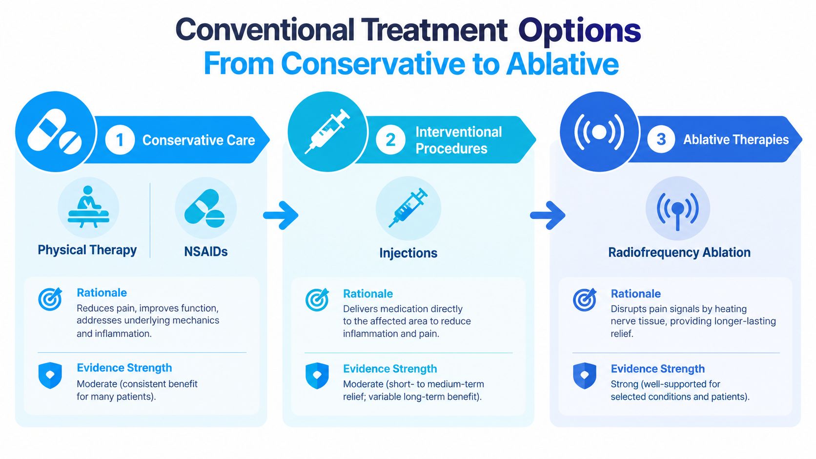
A patient usually moves through care in a fairly predictable order. First comes activity modification, physical therapy, manual therapy, and NSAIDs, especially when pain is mechanical and there are no red flags. The most helpful rehab programs usually focus on deep cervical flexor control and scapular stabilization, because those strategies reduce the burden on the neck during daily movement.
Interventions in the middle
If conservative care isn't enough, clinicians may use intra-articular injections or medial branch blocks with corticosteroid. These are usually time-limited tools, useful for diagnosis and sometimes for short-term symptom control. The effect is variable, and it doesn't rebuild the joint.
Radiofrequency ablation, or RFA, is the more durable conventional procedure. After positive controlled blocks, relief often lasts months rather than days, but the nerve eventually recovers and pain can return. For the right patient, that tradeoff makes sense.
Where the conventional ladder stops
Patients with facet cysts or structural instability sometimes need more specialized procedures, including surgical fusion in selected cases. The limitation is simple, conventional care can reduce pain and interrupt signaling, but it doesn't restore lost cartilage or reverse degeneration.
For clinicians comparing specialty workflows, a useful reference point is the PatientNotes orthopedics specialty, especially when you want a structured way to think about musculoskeletal care pathways.
Traditional treatment is strongest when the goal is symptom control. It's weaker when the patient wants tissue-level change.
Regenerative Injections and the Paracrine Mechanism
Regenerative injections sit in a different category from steroid shots and ablation. They use biologic materials such as stem cell preparations, PRP, or growth-factor concentrates to change the tissue environment around the cervical facet joint. The goal is biologic signaling, not immediate numbing.
What stem cells actually do
Stem cells do not mainly work by turning into new cartilage cells. Their benefit is described mostly through a paracrine mechanism, meaning they release soluble signals and extracellular vesicles that influence nearby cells rather than directly replacing damaged tissue (MSC review). A separate review of MSC paracrine effects explains that many therapeutic effects appear to come from soluble factors that shape local cellular responses (MSC paracrine review).
Those signals can help modulate inflammation, support repair, and influence the cells already living in the joint environment. In a mouse bone marrow model, direct in vivo evidence also showed MSC engraftment and differentiation over 10 weeks, including osteoblasts, osteocytes, and rare endothelial cells, while still treating paracrine signaling as a major mechanism.
For patients, the timing matters. Regenerative care is usually discussed in weeks and months, not minutes.
Why this matters for the facet joint
Facet pain is often confirmed with diagnostic blocks because imaging alone cannot show whether a joint is truly painful. False-positive blocks can also mislead treatment decisions, which is why the injection story matters as much as the scan. Regenerative injections try to treat the inflamed or degenerated joint environment that block testing has identified, rather than only silencing the nerve signal.
For a practical clinical example of biologic spine care, regenerative disc injections explained gives a useful frame for how these treatments are described. A related patient-focused overview of chronic pain care is available in how stem cell therapy works for chronic pain management.
Comparing Regenerative Therapy to Conventional Options
The cleanest way to compare options is by the kind of problem each one solves. Physical therapy addresses movement control, blocks help identify the pain source, steroid injections calm inflammation for a limited time, and RFA disables the pain-carrying nerve. Regenerative injections sit in a different lane, because they aim to support the joint environment itself.
Who tends to fit regenerative care
Younger patients with moderate arthropathy, patients who respond to diagnostic blocks but want to avoid repeated ablation, and people with multilevel disease often ask about regenerative therapy first. That interest makes sense when the goal is to preserve motion and avoid treating every painful level with a nerve-destructive approach. It also makes sense when repeat RFA starts to feel like a cycle instead of a plan.
Onset and durability are different too. Steroids may help faster, while regenerative effects usually build more gradually. Published regenerative outcomes for facet problems are still limited, so the evidence base is smaller than the conventional block-and-ablation pathway.
| Treatment | Onset | Durability | Best Candidate |
|---|---|---|---|
| Physical therapy | Gradual | Depends on adherence | Mechanical pain, deconditioning |
| Medial branch block | Immediate to short-term | Diagnostic, not durable | Confirming facet pain |
| Steroid injection | Days | Short-term | Inflammatory flare or bridge care |
| RFA | Weeks | Months | Block-positive facet pain |
| Regenerative injection | Weeks to months | Variable, limited data | Patients seeking tissue-level support |
The point isn't that regenerative care replaces every other option. It's that it may be a better fit for selected patients who want a biologic strategy instead of repeated nerve interruption.
What a Regenerative Treatment Journey Looks Like
A regenerative protocol starts with confirmation, not with the needle. Controlled medial branch blocks help identify the facet joint as the likely source, and then imaging and lab review make sure the plan is safe and anatomically sensible. That sequence keeps the procedure tied to a real pain pattern instead of a vague neck complaint.
What happens during the procedure
Some protocols use autologous bone marrow aspirate or adipose-derived cells, then concentrate and characterize the sample before injection. Others pair the biologic with PRP so there's both a cellular signal and a platelet-derived growth-factor signal. The injection is usually image-guided into the facet capsule and, when appropriate, around the nearby medial branches.
What patients usually monitor afterward
Recovery is measured over time, not in the recovery room. Patients often modify activity, follow a rehab plan, and come back for reassessment at intervals such as 6 weeks, 3 months, and 6 months. Staged treatment can make sense when disease is bilateral or spread across several levels.
For people comparing cervical and lumbar biologic care, stem cell neck pain treatment is a useful example of how neck-specific regenerative care is presented in practice.
Quick Reference for Patients and Clinicians
| Symptom Pattern | Likely Facet Levels | Recommended Action |
|---|---|---|
| Axial neck pain | C3-C4 through C6-C7 | Start conservative care, then consider medial branch blocks |
| Referred shoulder or scapular pain | Often C5-C6 or C6-C7 | Match the pain map, then test with targeted blocks |
| Whiplash-type pain | Upper and mid cervical levels | Watch for delayed mechanical pain and use controlled blocks |
| Rotation-limited pain | Any symptomatic cervical level | Assess extension-rotation loading, then choose the next diagnostic step |
A practical checklist helps keep decisions grounded. False-positive block risk should stay on the radar, dual-block confirmation matters before ablation, and regenerative therapy is most plausible when the patient wants tissue-supportive care rather than repeated denervation. The fastest way to get lost is to treat every painful neck the same way.
Frequently Asked Questions About Cervical Facet Joint Pain
Can an MRI confirm facet joint pain? No, MRI can show degeneration, but it can't prove that a cervical facet joint is the pain source. Structural change and pain don't always match.
What does a medial branch block do? It temporarily numbs the nerves that carry pain from a facet joint, which helps test whether that joint is driving the symptoms. Many clinicians use two blocks to lower the risk of a false-positive result.
Is radiofrequency ablation permanent? No, it's usually a months-long solution, not a permanent one, because the nerve can grow back over time.
How are stem cell injections different from PRP? PRP delivers platelet-derived growth factors, while stem cell-based approaches use living cells whose main benefit is paracrine signaling, not turning into new cartilage cells.
What does paracrine mean in plain language? It means the cells send out chemical messages that influence nearby tissue behavior, rather than replacing the tissue themselves.
Does whiplash facet pain heal on the same timeline as a simple strain? Not usually. Whiplash can irritate the joint capsule and supporting structures in a way that lasts longer than a muscle strain.
Are older patients with multilevel degeneration still candidates for regenerative therapy? Sometimes, yes, especially when the goal is to reduce irritation and support joint biology rather than chase a perfect scan.
Dream Body Clinic offers regenerative spine and joint care, including cervical-focused stem cell and PRP-based options for patients who want a biologic conversation about pain instead of only symptom suppression. If your neck pain pattern sounds facet-related, visit Dream Body Clinic to review evaluation and treatment options with a team that works in regenerative medicine.