
The most popular advice about stem cell treatments for rheumatoid arthritis is often the least useful: “They regenerate your damaged joints.” That explanation sounds reassuring, but it confuses tissue repair research with the way mesenchymal stem cells, or MSCs, are primarily known to work in rheumatoid arthritis.
MSCs don’t travel to an inflamed joint and turn into new cartilage, synovium, or bone. Their main proposed action is the paracrine effect, meaning they release specialized biological signals that influence nearby immune and joint cells. That distinction matters because a temporary reduction in inflammatory activity isn’t the same as durable disease control, permanent remission, or structural joint repair.
The Rheumatoid Arthritis Stem Cell Treatment at Dream Body Clinic is designed to go after the root cause of RA, an immune system that is attacking joints instead of protecting them.
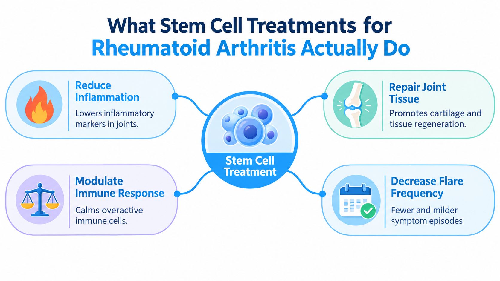
Table of Contents
- What Stem Cell Treatments for Rheumatoid Arthritis Do
- Why Paracrine Signaling and Immune Regulation Matter for RA
- Protocol Options, Cell Sources, and Dosing in Practice
- Routes, Symptom Effects, and the DMARD Comparison Question
- Safety, Candidacy, and the Evidence Patients Should Weigh
- Clinical Evidence Summaries From Published RA Trials
- Patient Journey, Monitoring, and What Realistic Outcomes Look Like
What Stem Cell Treatments for Rheumatoid Arthritis Do
Rheumatoid arthritis is driven by persistent immune activation. The immune system attacks joint tissues, especially the synovial lining, producing inflammation that can affect pain, swelling, movement, and long-term joint health. MSC treatment is therefore being studied as an attempt to recalibrate the inflammatory environment, rather than as a procedure that replaces worn joint parts.
The key mechanism is signaling
MSCs behave less like replacement bricks and more like temporary coordinators at a damaged construction site. They release a mixture of growth factors, cytokines, chemokines, microRNAs, and extracellular vesicles. Those signals can alter immune-cell behavior, influence inflammatory pathways, and support the body’s own repair responses.
The practical implication is important: infused MSCs are not expected to become new cartilage or rebuild a destroyed joint. Current reviews identify paracrine signaling and immunomodulation as their main proposed therapeutic value in rheumatoid arthritis. Reliable conversion into mature cartilage, bone, or synovial cells has not been established (International Journal of Molecular Sciences review).
What a meaningful response would look like
A response could include lower disease activity, fewer swollen joints, improved inflammatory blood markers, better function, or reduced stiffness. These changes would indicate that immune activity may have shifted. They would not, on their own, show that the autoimmune tendency has disappeared.
Patients need to separate four outcomes:
- Symptom improvement means you feel better.
- Immune modulation means inflammatory signaling may have changed.
- Disease control means objective activity stays suppressed over time.
- Structural repair means imaging or other evidence supports tissue restoration.
The categories can overlap, but they describe different results. A patient may feel relief early while the disease remains active or returns after the biological effect fades. That durability gap is one of the main questions to examine before treatment.
Practical rule: Judge treatment through objective measures collected over time, not through pain relief during the first few weeks or a testimonial alone.
MSCs are being investigated as temporary biological coordinators. Their signals can help reset inflammatory activity so that the body can heal.
Why Paracrine Signaling and Immune Regulation Matter for RA
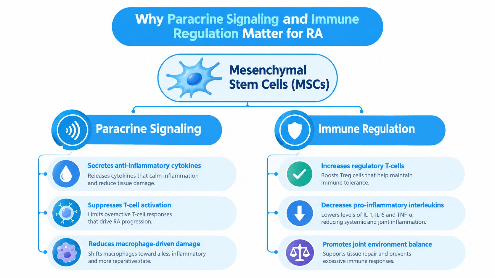
The immune system in rheumatoid arthritis involves several interacting cell types and inflammatory pathways. MSCs may influence that network through signals rather than by becoming replacement joint tissue.
Tregs, Th17 cells, and inflammatory balance
One important target is the relationship between regulatory T cells, or Tregs, and Th17 cells. Tregs help restrain excessive immune responses, while Th17-associated signaling is linked with autoimmune inflammation. Preclinical and clinical reviews describe MSC-related effects that may increase regulatory T-cell activity and reduce Th17-associated inflammatory signaling.
MSCs may also affect inflammatory mediators such as tumor necrosis factor-alpha and interleukin-6. Other reported signaling molecules include transforming growth factor-beta, indoleamine 2,3-dioxygenase, chemokines, growth factors, and extracellular vesicles. These mechanisms support a biological rationale for the Rheumatoid Arthritis Stem Cell Treatment.
Joint-lining cells and bone erosion
The target isn’t limited to circulating immune cells. Rheumatoid arthritis fibroblast-like synoviocytes are abnormal joint-lining cells that contribute to pannus formation and tissue destruction. MSC signals may inhibit their proliferation and invasive behavior.
The RANKL/OPG system is another relevant pathway because it helps regulate osteoclast activity, the process involved in inflammatory bone resorption. Influencing this system can matter for bone erosion.
Patients who want a broader introduction to immune-focused regenerative care can browse inflammation modulation formulas, while those comparing approaches for autoimmune disease can review this overview of stem cell therapy benefits for autoimmune conditions. Neither resource should replace a rheumatologist’s assessment or objective monitoring.
The most accurate summary is simple: MSCs can send signals that encourage a less destructive immune and joint environment. They don’t need to transform into cartilage or bone to influence those processes, and the clinical effect are outstanding.
Protocol Options, Cell Sources, and Dosing in Practice
“Stem cell treatment” describes a research approach, not one standardized procedure. Interpretation depends on the cell source, donor status, preparation, route, dose, infusion schedule, and medication used alongside it. Those variables can change both biological exposure and how long any improvement lasts.
Cell source changes the protocol
Published rheumatoid arthritis studies have examined MSCs from bone marrow, adipose tissue, and umbilical cord tissue. Autologous cells are collected from the patient, whereas allogeneic cells come from a donor. Autologous treatment requires collection and processing from the same person. Allogeneic treatment may simplify collection, but donor screening and manufacturing controls remain necessary.
For adipose-derived products, understanding fat-derived stem cells means examining how tissue is collected, processed, and characterized. The source label alone cannot show whether a product meets appropriate standards for sterility, viability, identity, or functional potency.
At Dream Body Clinic we have worked with Dr. Arnold Caplan, the man that named MSCs and he always recommended avoiding the use of the patients own MSCs for autoimmune diseases. This is because that persons MSCs are already compromised by their immune system. The best option is to get MSCs from someone else that is young and healthy which is why we use MSCs derived from Wharton Jelly (umbilical cord tissue) and placenta from live, healthy births. These young MSCs have not had enough exposure to an immune system to develop bad habits and heal autoimmune conditions the best – Rheumatoid Arthritis Stem Cell Treatment
What published dosing shows
One randomized, multicenter, double-blind, placebo-controlled dose-escalation phase Ib/IIa study enrolled 53 participants. It administered allogeneic adipose-tissue MSCs intravenously at 1, 2, or 4 million cells per kilogram, in three weekly injections. Researchers tracked DAS28-ESR, C-reactive protein, and ACR20 responses, as summarized in this published review.
A separate randomized dataset used umbilical-cord MSCs intravenously at 1 million cells per kilogram and followed participants for 48 weeks, with the protocol and cell-source context discussed in this comparison of perinatal umbilical cord tissue, placenta, and bone marrow stem cells. These figures describe particular research protocols, not a universal clinical dose.
Quality controls matter as much as the number
No universally accepted rheumatoid arthritis dose has been established. Before treatment, a clinic should specify:
- Manufacturing: Where and how the cells are processed.
- Sterility: How contamination is excluded.
- Viability: How many cells remain alive at administration.
- Potency: How the product’s intended biological activity is assessed.
- Route and schedule: Why intravenous or another route was selected.
- Rescue plan: What happens if disease activity worsens.
We provide all this information and more at Dream Body Clinic:
Routes, Symptom Effects, and the DMARD Comparison Question
The route of administration does not decide whether MSC therapy controls rheumatoid arthritis. It defines where the intervention is intended to act and what kind of result clinicians can reasonably evaluate.
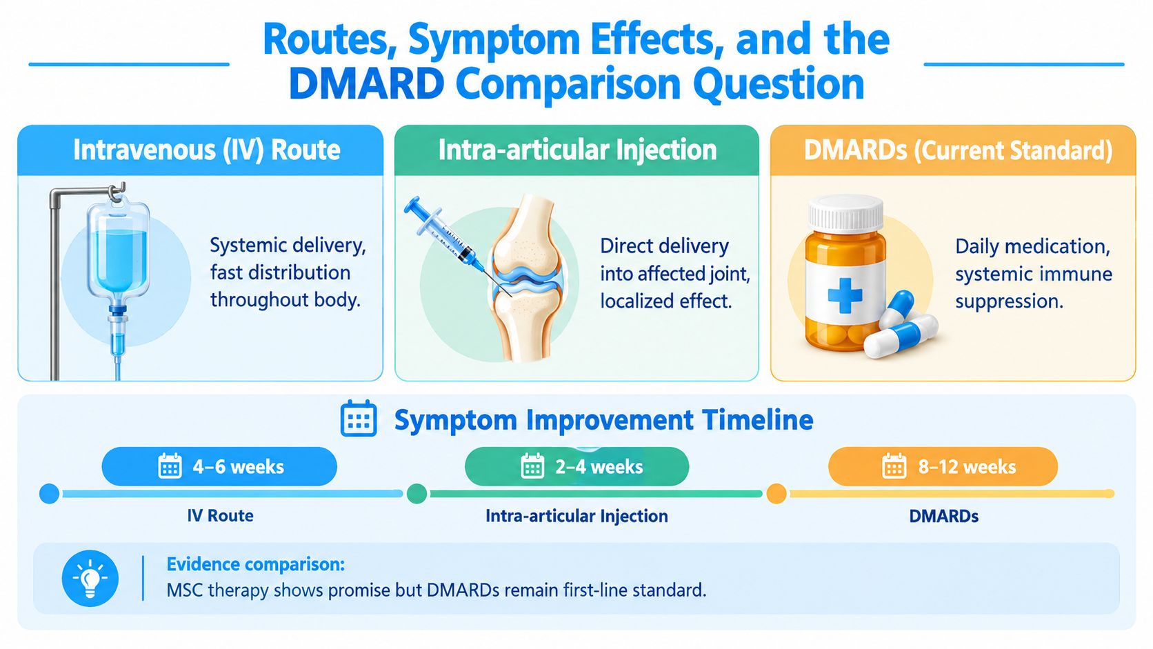
Published rheumatoid arthritis protocols have commonly used intravenous administration. Because RA involves immune activity throughout the body, an IV infusion is intended to expose circulating immune pathways to the cells and their paracrine signals.
What researchers have measured
Randomized studies have reported directional changes in DAS28, the Health Assessment Questionnaire, C-reactive protein, and erythrocyte sedimentation rate after MSC treatment. A systematic review of MSC therapy outcomes in rheumatoid arthritis described these findings as an efficacy trend requiring larger, higher-quality trials. The results therefore indicate a signal worth studying.
Route changes the clinical question. An intra-articular injection focuses on a particular joint, much like placing treatment at one address. An IV infusion is designed to influence broader immune activity. Neither approach should be assumed to rebuild every damaged tissue or remove the autoimmune process.

MSCs aren’t a proven replacement for DMARDs
DMARDs and biologic therapies remain established tools for controlling rheumatoid arthritis and limiting continuing inflammatory damage.
Early symptom improvement can be misleading. Reported benefits were not consistently maintained beyond 12 months when repeat treatment was not given. That durability gap is important because temporary immune recalibration can feel like disease control without demonstrating lasting change in the autoimmune process.
The studies are limited in what they can prove. At Dream Body Clinic we have over 9 years helping patients heal from Rheumatoid Arthritis symptoms with The Rheumatoid Arthritis Stem Cell Treatment and results have been amazing.
Safety, Candidacy, and the Evidence Patients Should Weigh
Safety for The Rheumatoid Arthritis Stem Cell Treatment is extremely high. In over 9 years we have never had an injury or infection from this treatment.

What pooled safety data show
A systematic review found no statistically significant difference in overall adverse events between MSC treatment and control. The pooled risk ratio was 2.35, with a 95% confidence interval of 0.58 to 9.58. Results also showed substantial heterogeneity, with I² approximately 59% (systematic review).
Studies are great tools to help validate what we know about the safety of this treatment, but over 9 years with no issues is much more important.
A conservative candidacy framework
A specialist assessment should document:
- Blood Analysis: We always ask patients to send recent bloodwork, but it is not required as our treatment includes a full blood panel
- Medication exposure: Record current and previous biologics, steroids, and treatment responses.
- Infection screening: Assess risks before an immunologically active intervention.
- Twelve-month follow-up: Monitor long enough to examine whether improvement persists.
The evidence base remains small. A 2025 review identified only 12 published clinical studies. Other reviews included studies involving 9 patients in a bone-marrow MSC study and 15 patients in an adipose-MSC phase I/IIa study. Many observation periods lasted no longer than six months (2025 review).
A temporary reduction in inflammation can be clinically meaningful, but it is not the same as sustained control of rheumatoid arthritis. We have patients from over 8 years ago that are still RA symptom free.
Clinical Evidence Summaries From Published RA Trials
Percentages from stem cell trials are easy to overread when dose, route, follow-up, and outcome definitions are removed. Each result needs its study design beside it. The central question is whether MSCs produced a temporary immune recalibration or durable disease control.
One randomized trial administered intravenous umbilical-cord MSCs at 1 million cells per kilogram. It included 52 participants in the MSC group and 53 participants in the control group, with follow-up to 48 weeks. A review of the umbilical-cord MSC randomized trial reported a total clinical effective rate of about 54% and stated that improvement in most patients lasted up to 12 months (review of the umbilical-cord MSC randomized trial).
A separate randomized phase Ib/IIa study enrolled 53 participants. It used allogeneic adipose-tissue MSCs intravenously at 1, 2, or 4 million cells per kilogram, delivered in three weekly injections. Reported ACR20 response rates in treated groups were approximately 20% to 45% after one month and 15% to 25% after three months, depending on dose. A review of the 53-participant adipose-MSC dose-escalation trial also reported a 29% response at one comparator time point and 0% at another (review of the 53-participant adipose-MSC dose-escalation trial).
| Study | Design | Cell Source | Dose and Route | Follow-up | Key Reported Outcome |
|---|---|---|---|---|---|
| Randomized umbilical-cord MSC trial | Randomized clinical dataset | Umbilical cord | 1 million cells per kilogram, intravenous | 48 weeks | About 54% total clinical effective rate, with improvement reported as lasting up to 12 months in most patients |
| Phase Ib/IIa dose-escalation study | Randomized, multicenter, double-blind, placebo-controlled | Allogeneic adipose tissue | 1, 2, or 4 million cells per kilogram, intravenous, weekly for three weeks | One and three months for reported ACR20 results | ACR20 responses varied by dose and time point |
| Autologous adipose pilot | Open-label, non-randomized phase I/IIa | Autologous adipose tissue | Intravenous | Week 52 | Improved tender and swollen joint scores, with no acute or long-term serious adverse events observed |
The autologous adipose pilot offers a useful safety observation, but its open-label, non-randomized design limits causal conclusions. It describes what happened in that group, not whether MSCs caused every improvement. Short-term paracrine effects may reduce inflammatory activity without proving that disease control will persist.
Now imagine how much better results can be with Allogeneic MSCs from the youngest, healthiest source possible – The Rheumatoid Arthritis Stem Cell Treatment
Patient Journey, Monitoring, and What Realistic Outcomes Look Like
A sensible patient journey starts with a clinical review, not a payment or infusion. The clinician should confirm the diagnosis, examine disease history and current medications, review previous responses, assess infection risks and organ health, and determine whether active rheumatoid arthritis is driving the symptoms.
Before treatment
Baseline records should include DAS28, CRP, ESR, swollen-joint counts, medication exposure, and infection screening. Imaging may help separate active inflammation from established structural damage. That distinction matters because reducing inflammation does not necessarily restore a joint that has already been damaged.
Ask the clinic:
- What cell source and preparation will be used?
- Is the product autologous or allogeneic?
- What sterility, viability, identity, and potency testing is performed?
- Why were this route and dose selected?
- Which medications will continue, pause, or change?
- Who will manage a flare or adverse event?
- Which objective outcomes will be reviewed at twelve months?
After treatment
The infusion may take only a few hours, but it provides no immediate proof of durable disease control. Structured follow-up at approximately three, six, and twelve months should assess symptoms, swollen-joint counts, CRP, ESR, DAS28, medication changes, and imaging when clinically appropriate.
A 2025 review of published RA MSC clinical studies reported that regulatory T-cell levels decreased between one and twelve months after treatment. Early symptom relief can therefore reflect a temporary paracrine effect rather than lasting control, making longer monitoring important.
Patients may also read about whether stem cell therapy can reduce rheumatoid arthritis pain. Pain reduction matters, but it does not by itself demonstrate remission, halted inflammation, or structural repair. The useful question is whether symptom changes are accompanied by objective findings that persist over time.
Realistic outcomes include reduction or elimination of inflammatory activity, improved function, fewer symptoms, and reduce or eliminated pain.
Dream Body Clinic offers individualized case reviews and regenerative-medicine protocols for international patients, including intravenous MSC-based approaches for autoimmune conditions. Patients considering an investigational plan can visit Dream Body Clinic with their medication history, laboratory results, imaging, and questions about durability, monitoring, and flare management.
The Rheumatoid Arthritis Stem Cell Treatment
Call (888) 704-3977 or WhatsApp +52 322 232 1055 for a Free Consultation