That sharp pop during a pickup game. The stubborn ache at the back of the heel that won’t settle down. The outer ankle pain that still feels “sprained” weeks later, except it also clicks, catches, or gives way on uneven ground. Many people in that moment search for one phrase, damaged tendons in ankle, hoping it means one simple problem with one simple fix.
It usually doesn’t.
That phrase can describe several different injuries in several different tendons. It might mean tendinopathy from overload, a partial tear, or a complete rupture. It might involve the Achilles tendon behind the ankle, the peroneal tendons along the outer ankle, or the posterior tibial tendon on the inner side that helps support the arch. Those distinctions matter because the right treatment for one can be the wrong treatment for another.
Heal an Injured Ankle fast with Stem Cell Therapy at Dream Body Clinic – Ankle Stem Cell Treatment – 50 Million Mesenchymal Stem Cells & and MRI
Table of Contents
- Introduction to Damaged Tendons in Ankle and What This Guide Covers
- How Ankle Tendons Work and Why They Get Damaged
- Recognizing Symptoms and Getting an Accurate Diagnosis
- Conservative Care and Early Loading Strategies That Protect Healing
- Surgical Repair Versus Regenerative Options and How Healing Signals Work
- Recovery Timelines Strength Deficits and What to Expect Month by Month
- Moving Forward With Confidence After Ankle Tendon Damage
Introduction to Damaged Tendons in Ankle and What This Guide Covers
A common mistake goes like this. Someone rolls an ankle, keeps walking, ices it for a few days, and assumes time will sort it out. But the pain doesn’t stay where a “typical sprain” should. Maybe it shifts to the outer ankle with a snapping feeling. Maybe push-off becomes weak. Maybe there was a clear pop in the calf and now rising onto the toes feels unreliable.
That’s when tendon involvement moves higher on the list.
Damaged tendons in ankle is an umbrella term, not a diagnosis. The ankle region contains several tendons with different jobs, different injury patterns, and different recovery paths. The Achilles handles powerful push-off. The peroneals help prevent the ankle from rolling. The posterior tibial tendon supports the arch and controls inward foot motion. When one of these structures is irritated, partially torn, or ruptured, the symptoms can overlap enough to confuse patients and even delay recognition.
One reason this topic deserves attention is that Achilles tendon rupture is not a rare edge case. It is described as the most common tendon rupture, and one review reported an annual incidence around 8 per 100,000 people per year, with published ranges across North America and Europe spanning roughly 2.5 to 47 per 100,000 person-years. That same review estimated a pooled global incidence of 15.7 per 100,000 person-years, rising from 6.1 in 1979 to 31.1 in 2021. In one long-term Finnish dataset, incidence increased from 2.1 per 100,000 person-years in 1979 to 21.5 in 2011. Achilles rupture also accounted for 10.7% of all tendon and ligament injuries in some datasets (PLOS One review on Achilles rupture epidemiology).
Why this matters: “Ankle tendon injury” isn’t one condition. The tendon involved and the stage of damage drive the next decision.
What usually helps most is a stepwise approach:
- Name the tendon: Back of heel, outer ankle, or inner ankle and arch.
- Name the injury stage: Overloaded tendon, partial tear, or complete rupture.
- Match treatment to stage: Protection, imaging, loading, surgery, or selected regenerative support.
By the end, you should be better able to describe your symptoms, understand why clinicians choose ultrasound or MRI, and know where conservative care ends and more advanced options begin.
How Ankle Tendons Work and Why They Get Damaged
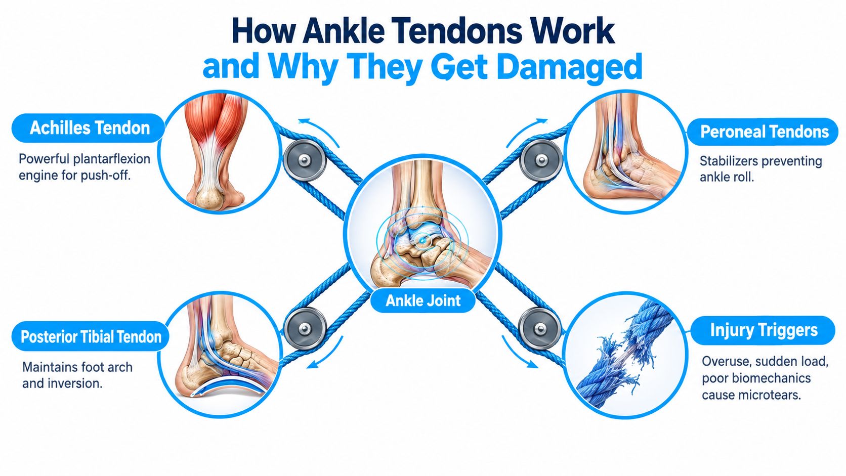
The ankle works a lot like a rope-and-pulley system. Muscles in the leg create force, tendons carry that force, and the ankle and foot bones act like pulleys that redirect it into movement. When the rope is healthy and the load is appropriate, motion feels smooth. When the rope is overloaded, frayed, or suddenly yanked, the system starts to fail.
The three tendons patients mix up most often
The Achilles tendon is the big power cable at the back of the ankle. It connects the calf muscles to the heel and drives plantarflexion, which is the motion used to push off, climb stairs, sprint, or rise onto the toes. When it’s irritated, people often feel stiffness and pain near the heel or in the tendon itself. When it ruptures, they often describe a sudden pop, loss of push-off strength, or the sense that someone kicked the back of the leg.
The peroneal tendons run along the outside of the ankle. Think of them as side guy-wires that steady the foot when the ground is uneven or the ankle starts to roll. They’re common troublemakers after a “sprain” that doesn’t behave like a routine sprain. People may feel lateral ankle pain, swelling, popping, catching, or pain on uneven ground.
The posterior tibial tendon runs along the inner ankle and helps support the arch. It acts like a sling under part of the foot. When it weakens, the arch may feel strained, the inner ankle may swell, and walking can become more tiring or awkward.
Tendons are hard to heal, but if just partially torn or hyperextended then Mesenchymal stem cells are an amazing way to fix them outside the traditional care options like surgery. Many people have avoided tendon surgery with this protocol – Ankle Stem Cell Treatment – 50 Million Mesenchymal Stem Cells & and MRI
How different kinds of damage happen
Not all tendon damage is a tear. That’s one of the biggest points of confusion.
A tendon can fail through overload and degeneration, often called tendinopathy. In that situation, the tendon is irritated and structurally stressed, but not fully torn. It can also fail through acute trauma, where a sudden force exceeds what the tissue can tolerate and causes a partial or complete tear.
A simple mental model helps:
| Pattern | What it usually feels like | What often caused it |
|---|---|---|
| Tendinopathy | Gradual pain, morning stiffness, soreness that warms up then returns | Repetitive overload, poor load progression |
| Partial tear | More focal pain, weakness, pain with push-off or resisted motion | Sudden overload on a vulnerable tendon |
| Complete rupture | Pop, sharp pain, immediate weakness or loss of function | High-force trauma or force through a degenerative tendon |
Tendon health is a balance between load capacity and load applied. Problems start when the demand outpaces what the tendon can currently handle.
Why location matters
If the pain is behind the heel, Achilles rises on the list. If it’s behind the outer ankle bone with snapping or catching, think peroneal. If it’s inside the ankle and into the arch, posterior tibial becomes more likely.
That’s why “rest it and see” can be misleading. Rest may calm pain for a few days, but it doesn’t tell you which rope in the pulley system is frayed.
Recognizing Symptoms and Getting an Accurate Diagnosis
A good tendon diagnosis usually starts with better language. “My ankle hurts” is too broad. “The outer ankle still pops after a sprain, especially on uneven ground” is far more useful. So is “I felt a pop in the calf and now I can’t push off normally.”
A large ankle MRI series in a football setting found tendon injuries in 37.0% of scanned ankles, and 31.6% of those tendon injuries involved the Achilles tendon. Within that Achilles group, 27.4% were tendinopathy and 4.2% were complete ruptures. Historical cohort data in the same review also noted that 70% of Achilles ruptures were sports-related, with male-to-female ratios in later reviews around 3.18:1 or 3.5:1, and peak incidence in middle age (review covering ankle tendon injury patterns and Achilles demographics).
Symptom clusters that point in different directions
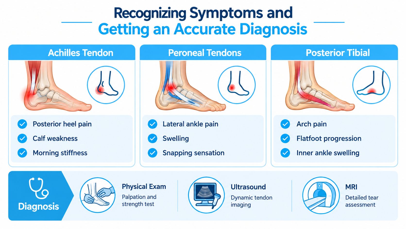
Here’s the practical pattern recognition clinicians use every day.
- Achilles pattern: Posterior heel or tendon pain, calf weakness, stiffness after rest, trouble with push-off.
- Peroneal pattern: Outer ankle pain after a sprain, swelling behind the fibula, popping, catching, or pain on uneven ground.
- Posterior tibial pattern: Inner ankle pain, arch discomfort, swelling along the inside of the ankle, sense that the foot is flattening or tiring.
If your symptoms include a shoulder-like pop or snapping sensation in another tendon region, the same principle applies. People often find it helpful to compare how tendon mechanics produce odd noises and catching sensations in other joints, such as this overview of Ankle Stem Cell Treatment – 50 Million Mesenchymal Stem Cells & and MRI
Exam, ultrasound, and MRI each answer different questions
Physical examination still matters. A clinician checks tenderness, swelling, strength, push-off, tendon continuity, and whether the symptoms can be reproduced with motion or resisted testing. But tendon injuries are often underrecognized early, especially when swelling from a sprain clouds the picture.
For peroneal tendon injuries, imaging choice changes what you catch. In one comparative study, ultrasound showed 88% sensitivity and 100% specificity for tendon tears, while MRI reached 100% sensitivity and specificity. The same study found ultrasound was better for tendinopathy and subluxation, while MRI was slightly better for tears (comparative ultrasound and MRI study for peroneal tendon pathology).
That leads to a sensible real-world workflow:
- Dynamic symptoms first: If the tendon snaps, slides, or hurts during motion, ultrasound can be very helpful because it watches the tendon move in real time.
- Suspected tear or uncertain extent: MRI gives more detail when a tear is suspected or the treatment decision depends on showing how much tissue is disrupted.
- Need to separate partial from complete rupture: That distinction matters because the recovery plan changes substantially.
If a clinician can’t yet tell whether you have tendinopathy, a partial tear, or a complete rupture, imaging isn’t just confirming the diagnosis. It’s deciding the treatment lane.
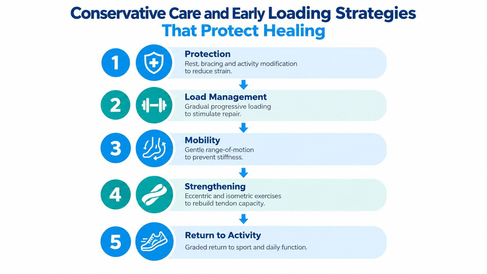
Conservative Care and Early Loading Strategies That Protect Healing
People often hear two pieces of advice that sound contradictory: protect the tendon, and start moving early. Both can be correct. The trick is matching the amount of protection to the type and stage of injury.
For Achilles rupture, delayed or poorly positioned immobilization can increase the risk of functional lengthening, where the tendon heals too long and push-off stays weak. Expert guidance recommends immediate plantarflexed immobilization with non-weight-bearing, and non-operative pathways often begin within 48 hours, then transition to an Achilles boot with heel lifts at about 2 weeks. When the extent of injury is unclear, ultrasound or MRI is commonly used to confirm whether the rupture is partial or complete (AJR review on Achilles rupture imaging and immobilization principles).
The care ladder that makes sense
Most non-surgical care works best as a progression, not a random pile of treatments.
-
Protect what’s acutely vulnerable
A suspected rupture or clear high-grade tear needs immediate protection. That can mean plantarflexed positioning, a boot, crutches, and reducing strain before the tendon stretches out while trying to heal. -
Settle symptoms without going completely idle
Tendons hate chaotic overload, but they also don’t love prolonged underuse. Activity modification helps. So does choosing movements that don’t provoke sharp pain or instability. -
Reintroduce motion carefully
Once the tendon is protected enough, gentle range of motion helps prevent a stiff, guarded ankle. The exact timing depends on the tendon and whether there’s a tear.
Before moving deeper into exercise progressions, some readers like a practical explanation of what to expect with mesenchymal stem cell therapy, because it sometimes comes up as an adjunct option for tendon pain rather than a stand-alone fix.
Early loading is not the same as rushing
Many generic guides lose people. Early loading doesn’t mean “walk it off.” It means adding force in a way the healing tissue can tolerate.
A useful progression often includes:
- Isometrics first: Static calf or foot loading to wake the system up without much tendon excursion.
- Controlled calf work next: Gradual heel raise progression, seated before standing if needed.
- Kinetic-chain work: Hip strength, balance, and movement quality. Tendons don’t function in isolation.
- Terrain and gait retraining: Especially for peroneal symptoms and for anyone who still feels unstable on uneven ground.
This can be easier to follow with visual coaching, especially when trying to pace activity day by day.
For practical daily pacing, a structured resource such as these activity recommendations for orthopedic recovery can help patients avoid the common cycle of doing too much on a good day and paying for it the next two days.
Practical rule: If pain, swelling, or limping clearly escalates the day after loading, the tissue is telling you the dose was too high.
Red flags for escalation include loss of push-off, obvious deformity, recurrent snapping, persistent giving-way, or a plateau despite well-structured rehab.
Surgical Repair Versus Regenerative Options and How Healing Signals Work
When tendon damage is severe, the conversation shifts from “How do we calm this down?” to “What is the tendon physically able to do right now?” That’s the point where surgery and regenerative options often enter the discussion together.
What surgery aims to fix
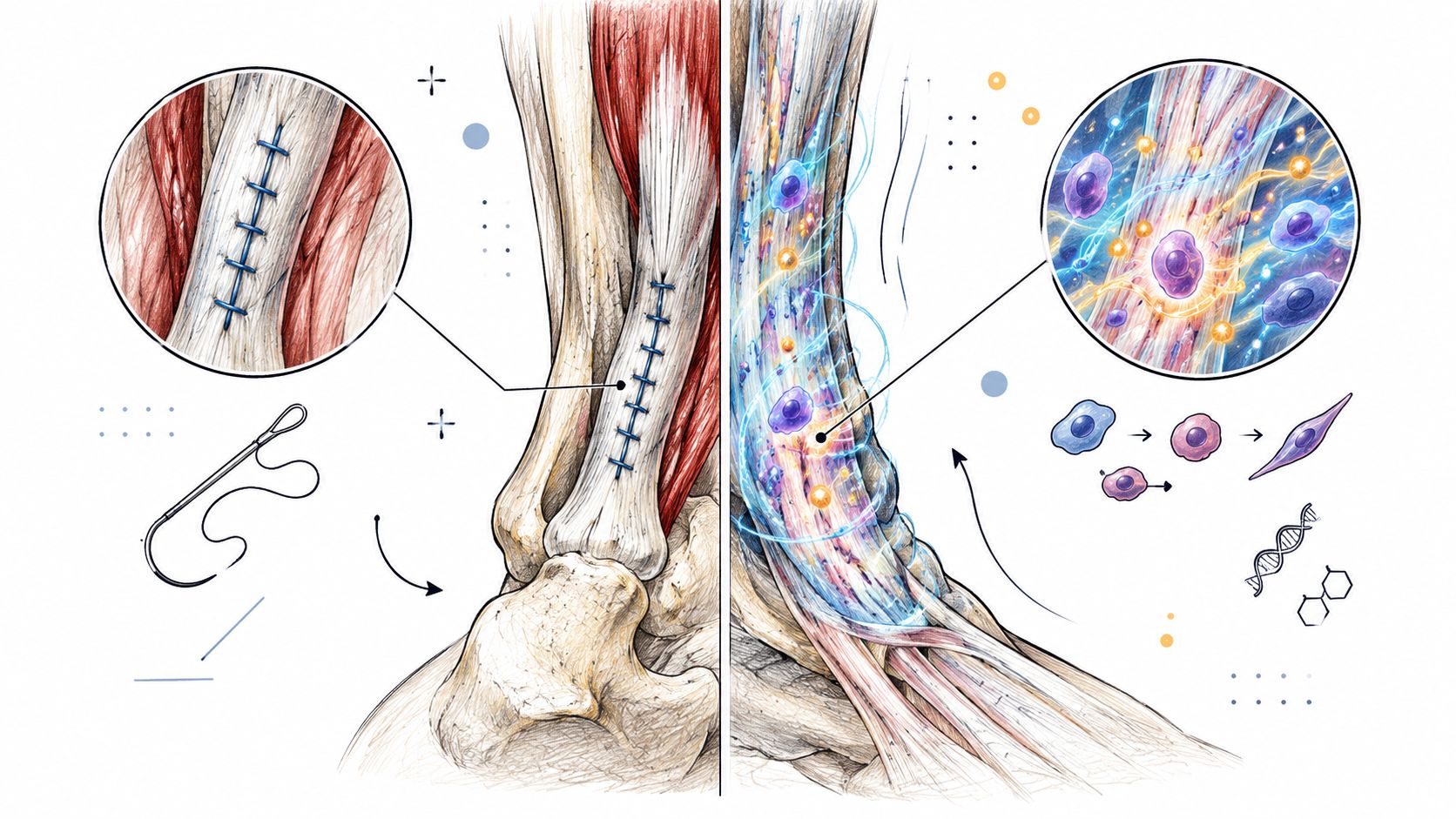
Surgical repair is typically considered when there is a complete rupture, a tear that doesn’t functionally improve, recurrent tendon instability, or tissue quality that won’t support recovery with conservative care alone. The surgical goal is mechanical. Reconnect or stabilize damaged tissue, restore tendon tension, and improve the tendon’s ability to transmit force.
That mechanical reset can be very helpful, but it comes with downtime, staged rehabilitation, and the need to protect the repair while it matures. If you’re comparing options, it helps to understand the practical milestones in Achilles tendon surgery recovery so expectations stay realistic.
What regenerative care is actually trying to do
Regenerative approaches ask a different question. Instead of mechanically stitching tissue back together, they try to support the healing environment.
A point needs to be stated plainly. Mesenchymal stem cells do not work by differentiating into replacement tendon cells in a meaningful long-term way. They work mainly through the paracrine effect, which means they send out specialized signals, including bioactive factors and exosomes, that help guide the healing process. A 2021 review states that accumulating evidence indicates stem cells promote tissue healing mainly via paracrine mechanisms, with effects tied to reduced inflammation, less fibrosis, and improved regeneration through signals that influence host cells (review on MSC paracrine mechanisms in tissue healing).
Heal Your ankle fast with Ankle Stem Cell Treatment – 50 Million Mesenchymal Stem Cells & and MRI
A balanced way to compare the two
| Option | Main goal | Best fit |
|---|---|---|
| Surgical repair | Restore continuity, tension, and stability | Complete rupture, unstable tear, failed conservative care |
| Regenerative support | Influence healing through signaling | Selected partial tears, chronic tendon pathology, adjunct use after surgery in some pathways |
There is also ankle-region context for these therapies. A 2022 orthopedic review on ankle osteochondral lesions and ankle osteoarthritis notes that mesenchymal stem cells are being used in the ankle either alone or after surgery, which shows that ankle-region cell therapy is already being discussed within structured treatment pathways in published literature (ankle orthopedic review discussing MSC use alone or after surgery).
One clinical option in this space is stem cell ankle treatment, which is presented as an ankle-focused regenerative pathway rather than a replacement for proper imaging, diagnosis, or surgical decision-making when a rupture needs mechanical repair.
Recovery Timelines Strength Deficits and What to Expect Month by Month
Recovery doesn’t move in a straight line. You improve in layers. Pain settles first, then walking normalizes, then strength starts to return, and only later do confidence and higher-demand tasks catch up.
One under-discussed point is immobilization length. A 2025 analysis reported that ankle-function recovery was better when immobilization lasted 0 to 2 weeks compared with longer protocols, with better range of motion, strength, and patient satisfaction and no significant rise in complications or re-rupture. The same body of emerging material also reinforces that heavy, progressive calf loading at roughly 70 to 90% 1RM over at least 12 weeks is central in Achilles tendinopathy care, and that insertional cases may need different loading angles than midportion cases (review discussing immobilization duration and progressive loading in Achilles care).
A realistic month-by-month view
Early phase
The focus is protection, settling irritation, and beginning safe motion when the tendon allows. Walking often still looks guarded. If you’re temporarily non-weight-bearing, practical equipment matters. This guide to using a knee scooter can make everyday mobility less clumsy while the ankle is protected.
Building phase
People often get impatient. Pain may be lower, but the tendon still isn’t ready for explosive loading. Calf strength, single-leg control, and ankle mobility need steady rebuilding.
Higher-function phase
Later rehab is less about symptom relief and more about restoring performance. That means push-off, balance, uneven-ground confidence, and tendon tolerance to repeated loading.
Recovery is more than pain reduction
A 2025 systematic review found that Achilles tendinopathy is associated with measurable deficits in ankle dorsiflexion, plantarflexion endurance, hip extension strength, hop performance, and foot alignment (systematic review on broader deficits in Achilles tendinopathy).
That’s why a rehab plan aimed only at “making the tendon less sore” often falls short.
- Dorsiflexion matters: If ankle motion stays restricted, gait and loading patterns stay altered.
- Calf endurance matters: The tendon may tolerate one good repetition but fail during repeated steps, hills, or sport.
- Hip strength matters: Weakness upstream changes how force travels down the leg.
- Hop and uneven-ground control matter: Many patients feel “mostly fine” until the surface becomes unpredictable.
Better walking is an early milestone. Better force production is a later one. Those are not the same recovery marker.
Moving Forward With Confidence After Ankle Tendon Damage
The clearest way to think about damaged tendons in ankle is to hold three questions in mind.
First, which tendon is involved. Back of the heel suggests Achilles. Outer ankle popping or catching points more toward peroneals. Inner ankle and arch symptoms raise posterior tibial concern.
Second, what stage of damage is present. An overloaded tendon behaves differently from a partial tear, and both differ sharply from a complete rupture.
Third, which care path matches that stage. Some injuries need immediate protection and imaging. Others need a disciplined loading plan. Some require a surgical opinion. Some may be candidates for regenerative support that works through signaling rather than cell replacement.
A short checklist helps before your next appointment:
- Track the exact location: Back, outer side, or inner side of the ankle.
- Note the trigger: Gradual overload, sudden twist, jump, sprint, or a felt pop.
- Describe function loss: Trouble pushing off, going downstairs, balancing, or walking on uneven ground.
- Watch for mechanical symptoms: Snapping, catching, giving-way, or visible change in contour.
- Bring prior imaging if you have it: It saves time and improves decision-making.
Most tendon injuries do improve when the diagnosis is accurate and the load progression is sensible. What delays progress is usually not bad luck. It’s treating the wrong tendon, the wrong injury stage, or the wrong amount of protection.
If pain, weakness, or instability persists, get a specific evaluation rather than broader reassurance.
To avoid surgery and heal your ankle fast check out Ankle Stem Cell Treatment – 50 Million Mesenchymal Stem Cells & and MRI
Dream Body Clinic offers orthopedic regenerative care that includes ankle-focused MSC treatment pathways for patients dealing with tendon-related pain, partial tears, or chronic ankle problems, along with imaging-based planning where appropriate. If you want to explore whether a regenerative approach fits your diagnosis and stage of injury, visit Dream Body Clinic.
Call us at (888) 704-3977 or on WhatsApp at +523222321055 for a free consultation