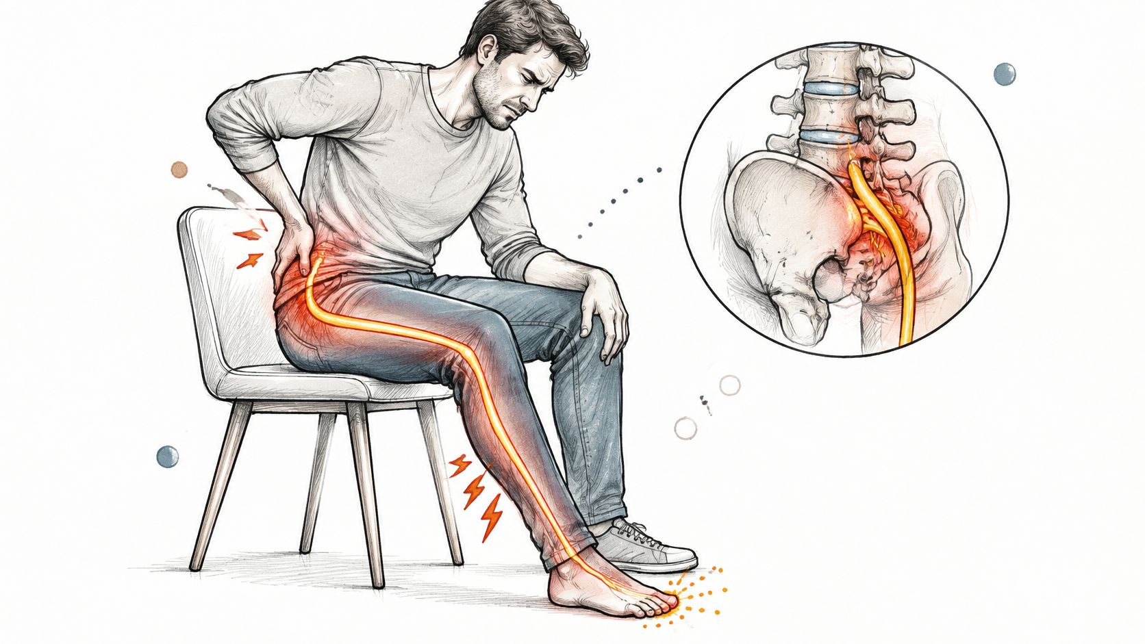

You've had an aching lower back for years, but now the pain runs down your leg. Your MRI report mentions degeneration, a bulge, and nerve compression, while different clinics recommend exercise, injections, surgery, or stem cells. It's hard to know which finding matters and which treatment actually fits.
Degenerative disc disease and sciatica aren't interchangeable diagnoses. Understanding the connection starts with identifying the affected nerve, checking how well it works, and deciding whether treatment should focus on pain control, function, or urgent protection of neurological function.
Table of Contents
- How Disc Degeneration Turns Into Sciatica
- Symptoms That Signal Nerve Involvement and Red Flags
- Conservative and Interventional Treatment Pathways Compared
- Diagnosis Workflow and the Pathway to Follow
- How MSC Therapy Works Through Paracrine Signals
- Who Qualifies for Regenerative Therapy for Sciatica
- What Results to Expect and When
How Disc Degeneration Turns Into Sciatica
Consider an illustrative example: a 48-year-old warehouse worker has managed backache for years, then wakes with burning pain running from the buttock to the foot. The important change isn't merely stronger pain. It's a new pattern suggesting nerve involvement.
A spinal disc acts like a shock absorber, with a jelly-like center inside a tougher outer ring. During degeneration, the disc loses water and cushioning molecules, the ring can develop fissures, and disc height may decrease. Disc material can then bulge or herniate, rather like a worn gasket pressing against a nearby wire.
The diagram shows how structural changes can become nerve symptoms.
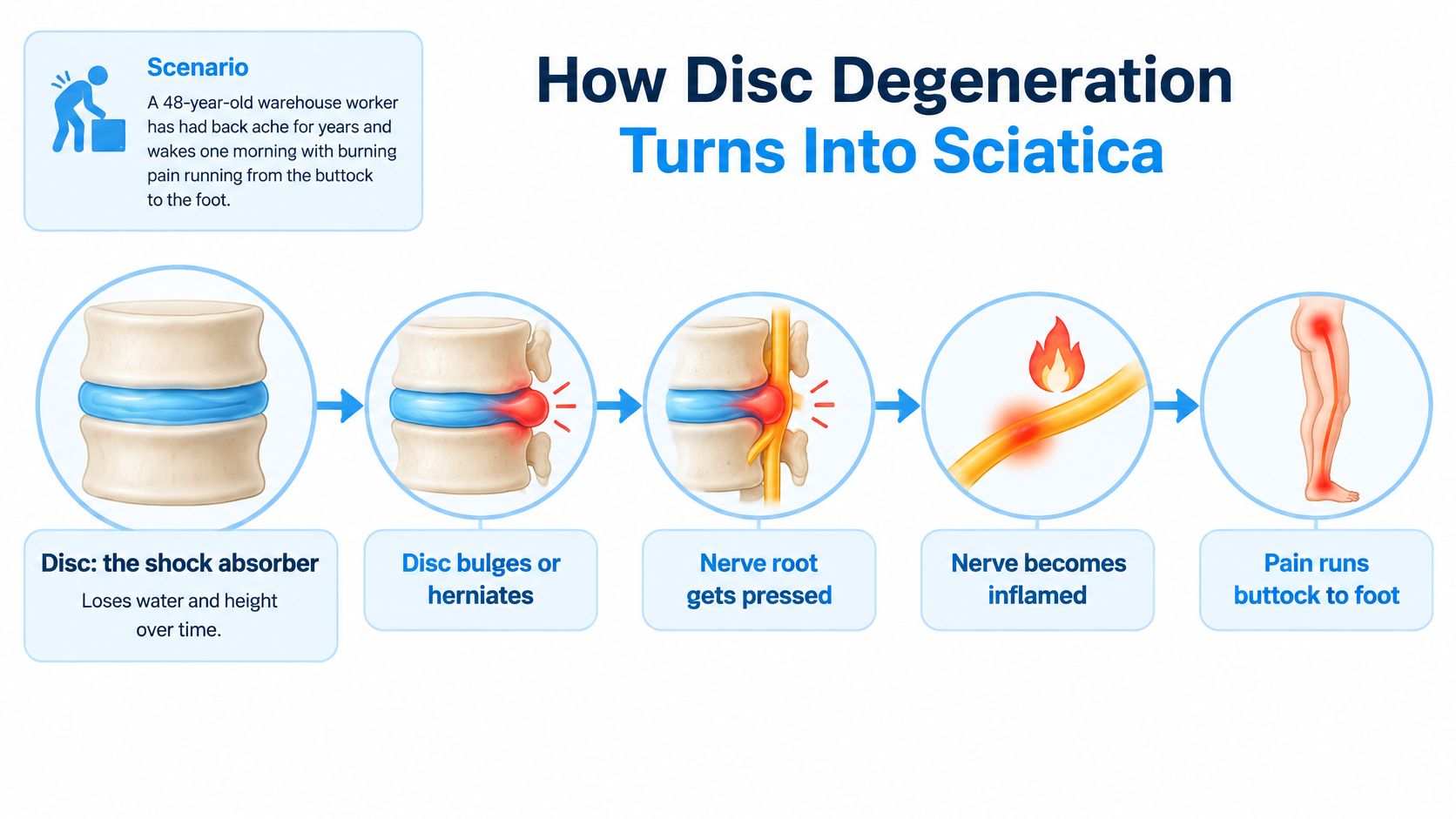
From a disc finding to a nerve pattern
Sciatica develops when disc material, inflammatory chemicals, or both affect a lumbosacral nerve root. That helps explain burning, electric pain, tingling, and sometimes weakness, rather than just an ache in the back.
Slightly more than 90% of lumbar disc herniations occur at L4-L5 or L5-S1, leaving fewer than one in ten elsewhere, according to the clinical overview of sciatica and lumbar nerve-root involvement. The following patterns are typical, not universal, because the position of a herniation also matters.
Disc level, nerve root and the symptoms each one produces
| Disc level | Nerve root affected | Typical pain route | Exam finding to check |
|---|---|---|---|
| L4-L5 | Usually L5 root | Buttock and outer thigh to the top of the foot and big toe | Weak big-toe extension, reduced sensation on the top of the foot |
| L5-S1 | Usually S1 root | Back of the thigh and calf to the heel and sole | Weak push-off on the toes, reduced Achilles reflex |
| L3-L4, less common | Usually L4 root | Front of the thigh down the inner shin | Weak knee extension, reduced knee-jerk reflex |
Clinicians connect a level to a leg by examining muscle strength, sensation in a defined territory, and reflexes. Not every affected nerve produces all these findings, and pain location alone can't establish the diagnosis.
For related terminology, compare this explanation of degenerative disc disease and spondylosis. For another patient-facing discussion of degeneration and narrowing around nerves, the disc degeneration insights from Aspen Falls Wellness offer useful background.
Symptoms That Signal Nerve Involvement and Red Flags
Mechanical back pain often feels like an ache linked to bending, lifting, or sitting. It may remain around the lower back and buttock, whereas nerve-root pain more often follows a recognizable route down the leg and can extend below the knee.
Coughing or sneezing may aggravate radiating pain. A strip of numbness, persistent pins and needles, or a foot that catches while walking deserves particular attention because these symptoms may reflect more than pain sensitivity.
Low back pain ranks first globally for disability burden, according to the Global Burden of Disease analysis of lumbar pain and disability, which helps explain why persistent leg symptoms deserve assessment.
Warning signs that shouldn't wait
Seek same-day medical assessment for the following changes. New bladder or bowel dysfunction, saddle numbness, or rapidly worsening weakness warrant emergency evaluation rather than a routine appointment.
- Progressive leg weakness: Your foot increasingly drags, your knee gives way, or strength is clearly deteriorating.
- New bladder or bowel changes: Difficulty emptying the bladder, loss of control, or an unexplained change accompanying back and leg symptoms.
- Saddle-area numbness: Reduced feeling around the groin, genitals, inner thighs, or buttocks.
- Sudden severe pain after trauma: Especially when accompanied by difficulty standing, walking, or new neurological symptoms.
These signs can change the decision from a planned rehabilitation programme to urgent spinal assessment, including surgical review when necessary. Don't use an exercise trial or regenerative consultation to postpone that assessment.
What to record before your appointment
Bring a short symptom history rather than trying to diagnose yourself. Record when the leg pain began, where it travels, what aggravates it, and any change in strength or sensation.
Include disrupted sleep and night pain, but don't assume night pain alone proves nerve compression. Home observations help a clinician assess you; they don't replace a neurological examination.
Conservative and Interventional Treatment Pathways Compared
A patient with stable leg symptoms may start with guided exercise, while someone with worsening weakness needs prompt surgical assessment. Treatment depends on how symptoms, nerve findings, and imaging fit together. Reassess each step rather than treating options as a checklist.
A 2025 systematic review of 50 randomized trials involving 4,920 participants reported short-term leg-pain reductions with some approaches, including spinal manipulation and exercise combined with neural mobilization. The certainty was very low. The review did not establish high-quality evidence that any non-surgical intervention is superior for chronic sciatica.
Compare purpose before promises
Sciatica treatment options by suitability, evidence and benefit window
| Option | Best suited for | Evidence and certainty | Typical benefit window |
|---|---|---|---|
| Structured exercise with neural mobilization | First-line care for stable radicular symptoms | Some trials found short-term leg-pain improvement; very low certainty | Short-term response, reassessed during rehabilitation |
| Spinal manipulation | Selected patients seeking hands-on care alongside exercise | Some trials found short-term leg-pain improvement; very low certainty | Short term |
| Oral medication, including anti-inflammatories and nerve-pain agents | Selected symptom flares after medication review | Benefits and harms vary by drug; choices are not interchangeable | Short-term use with review |
| Epidural steroid injection | Selected radicular pain with clinical and imaging agreement | Aims to relieve symptoms, not rebuild the disc; benefit can be temporary | Variable, often temporary |
| Surgical decompression, such as microdiscectomy | Significant compression with deterioration, or persistent symptoms despite appropriate care | Requires agreement among symptoms, examination, and imaging; comparative outcomes vary | Judged through neurological recovery and function |
| MSC injection, investigational | Carefully evaluated patients considering an emerging option | Clinical benefit remains uncertain; stronger randomized trials are needed | No dependable benefit window established |
Medication calls for an individual review. Ask which symptom a prescription targets, what side effects to watch for, and when to stop if it does not help.
An epidural injection targets symptoms around an irritated nerve. It does not repair the disc. Ongoing pain alone does not show that repeated injections are appropriate.
Decompression isn't the same as fusion
Microdiscectomy removes disc material that is irritating or compressing a nerve. Fusion joins spinal segments, so evidence about one procedure cannot establish the value of the other. Trials have compared fusion with non-operative care for lumbar degenerative conditions, with mixed results. That comparison does not determine which procedure suits an individual.
Ask the surgeon to identify the anatomical target and explain the functional improvement they expect. The comparison of herniated-disc treatment options can help you prepare questions for that discussion.
Apply the same checks to any proposed treatment, including regenerative options: does the suspected disc level match the nerve-root symptoms and examination findings, what outcome is expected, and how certain is the evidence? Track success through walking, sleep, medication use, strength, and neurological symptoms, not a promise that the disc will heal.
Diagnosis Workflow and the Pathway to Follow
A patient may hold a dramatic MRI report while describing only mild leg symptoms. The useful question is whether the scan explains the symptoms, not whether the report sounds serious.
A scan is a map, not a verdict. Disc degeneration, bulges, and joint changes can occur without symptoms, so identifying an abnormality doesn't establish that it causes your pain.
This flowchart summarizes the clinical sequence.
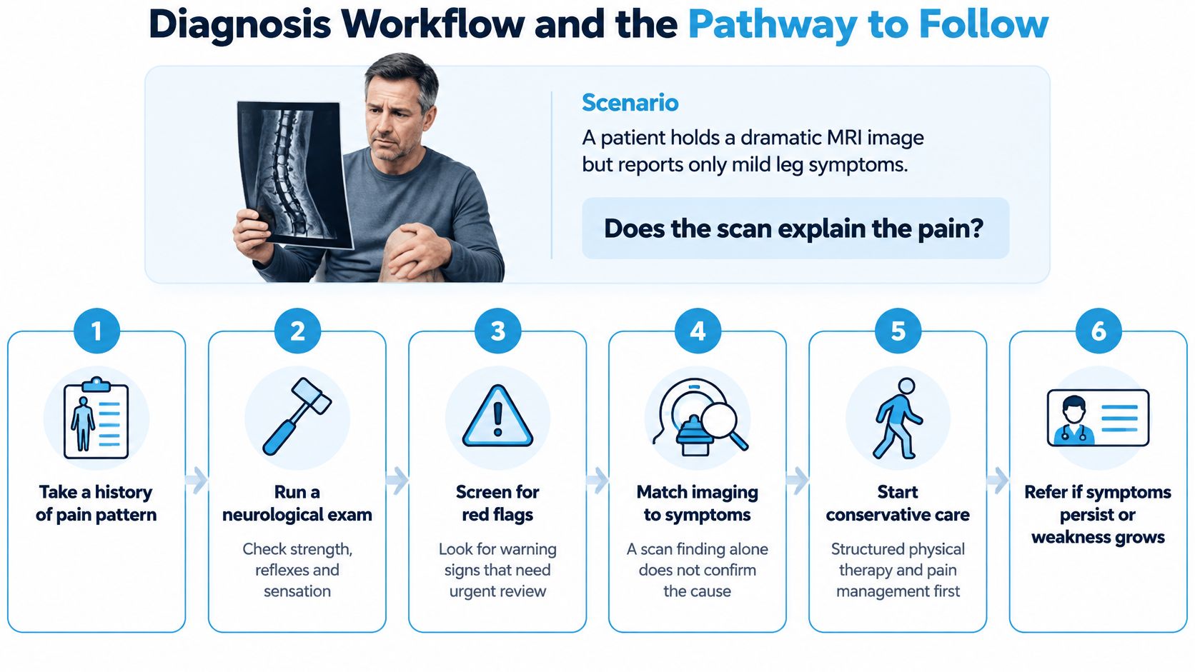
Establish the diagnosis before choosing a procedure
Describe the pattern and timeline. Explain where symptoms begin, where they end, whether back or leg pain dominates, and how daily activities have changed. A body diagram can be more useful than a long list of pain adjectives.
Document the neurological examination. The clinician should record myotomal strength, meaning muscles supplied mainly by a particular root; dermatomal sensation, meaning its skin territory; and reflex differences. Ask which findings support the suspected level.
Screen for urgent warning signs. The red flags above take priority over routine treatment planning. A concerning change should trigger urgent assessment rather than another appointment in a standard programme.
Use MRI when it can change management. NICE guidance on low back pain and sciatica recommends against routine imaging in non-specialist settings. Specialist imaging is appropriate when its result is likely to influence care, such as evaluation of significant deficits, suspected serious pathology, or an invasive treatment decision.
Apply the concordance test. The scan's side and level should fit the pain route and examination. For example, a finding on the opposite side deserves an explanation before anyone treats it as the cause.
Move from diagnosis to a monitored plan
Complete structured care with explicit goals. Agree on activities you want to regain and a review point. Repeat examinations help distinguish ongoing pain with stable function from worsening nerve dysfunction.
Escalate for a reason. Persistent disability despite appropriate treatment may justify an injection discussion or surgical review. Ask what new information supports escalation and what would make the clinician reconsider.
Scrutinize regenerative protocols. Request the published protocol, cell dose, delivery route, relevant clinical evidence, exclusion criteria, and follow-up plan. A study of local disc treatment doesn't automatically support a different route or a combined treatment package.
Practical rule: Before agreeing to an invasive treatment, ask the clinician to show how your symptoms, examination, and imaging identify the same target.
Evidence comparing surgery with conservative treatment is mixed, so neither an alarming scan nor persistent pain should substitute for individual assessment. Neurological deterioration changes the urgency; otherwise, the discussion should include alternatives and uncertainty.
For an additional explanation of lower-back regenerative care, the following video may support questions for a consultation.
Bring prior imaging, medication history, and a written account of functional and sensory changes. For regenerative treatment, the decision should rest on your clinical pattern, progress, and risks, not enthusiasm about an emerging technology.
How MSC Therapy Works Through Paracrine Signals
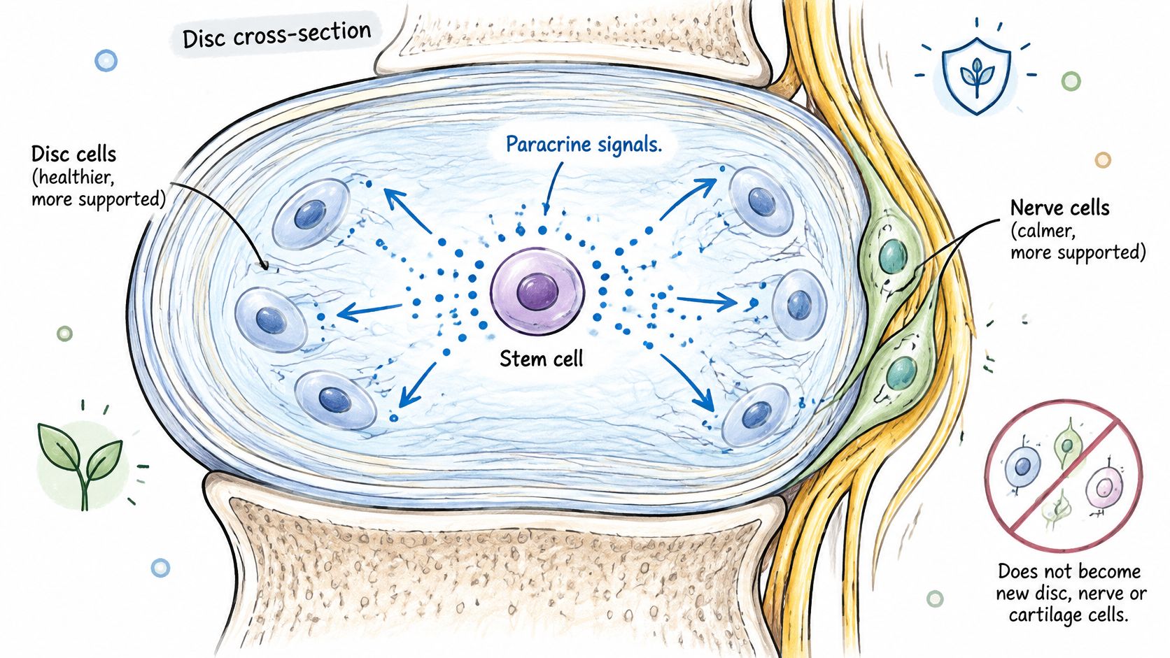
Mesenchymal stromal cells, commonly called mesenchymal stem cells or MSCs, don't work by differentiating into replacement disc, nerve, or cartilage cells in the patient. Describing them as building a new disc misrepresents the proposed mechanism.
A review of MSC mechanisms in cardiovascular research found no clear, consistent in-vivo or clinical evidence of tissue-specific differentiation. Implanted bone-marrow MSCs also showed limited persistence, often lasting no longer than about three weeks. Those findings concern cardiovascular research, not proof of how every spinal injection behaves.
Signals rather than replacement parts
The main explanation is the paracrine effect. MSCs release biologically active messages that influence neighboring cells and the local tissue environment.
Think of a site foreman radioing instructions to workers already on the job. The foreman doesn't become the building materials; similarly, MSCs may influence the patient's existing cells rather than becoming replacement tissue.
MSCs work by sending out specialized signals to guide the healing process, not by turning into new disc or nerve cells.
The illustration represents that signaling relationship.
The released mixture is called the secretome. It includes cytokines, chemokines, growth factors, extracellular vesicles, and microRNAs, as described in research on the MSC secretome and paracrine activity.
Specific signals identified in research include VEGF, HGF, IGF-1, SDF-1 alpha, TGF-beta, and TSG-6. These participate in overlapping pathways involving cell survival, inflammation, blood-vessel formation, and resident repair cells, according to this review of MSC-derived signaling factors.
What the mechanism does and doesn't establish
Researchers associate secretome activity with several effects:
- Inflammatory regulation: Changes in inflammatory signaling.
- Immune-cell behavior: Macrophage shifts toward pro-regenerative activity.
- Vascular responses: Promotion of blood-vessel formation in studied settings.
- Fibrosis modulation: Effects on scarring-related pathways.
These effects are discussed in research on MSC secretome-mediated repair. They provide biological plausibility, not proof that a particular injection relieves sciatica or reverses degeneration.
The explanation of how stem cell injections work can support a mechanism-focused consultation. For patients, the appropriate expectation is a possible signaling effect on inflammation and local repair, not a rebuilt disc or guaranteed release of mechanical nerve compression.
Who Qualifies for Regenerative Therapy for Sciatica
Consider a hypothetical 55-year-old office manager with six months of leg pain, stable strength, and an L5-S1 finding that matches an S1 symptom pattern. Exercise and medication haven't restored function despite two structured treatment attempts.
That profile is more appropriate for a careful investigational-treatment discussion than a patient whose strength is deteriorating. It still doesn't prove that MSC treatment will help or establish formal eligibility.
Selection means checking the whole pattern
Patient selection checkpoints for regenerative sciatica care
| Checkpoint | Favours a regenerative option | Favours deferring or ruling out |
|---|---|---|
| Symptom duration | Persistent for months despite structured care | Recent onset and improving |
| Neurological examination | Stable strength and sensation without progression | Progressive weakness or cauda equina features |
| Imaging concordance | Findings match the side and route of leg symptoms | Opposite-side abnormality or poor symptom agreement |
| Prior conservative care | Completed exercise, medication review, and possibly an appropriate injection | Basic care hasn't been tried |
| Pain pattern | Discogenic or radicular pattern identified clinically | Possible hip, sacroiliac, or peripheral-nerve cause |
| Expectations | Goals based on walking, sleep, and function | Expectation of a normal-looking disc on follow-up MRI |
| Systemic health | Conditions disclosed and cleared by a physician | Active infection, malignancy concerns, or uncontrolled systemic disease |
These are discussion checkpoints, not a validated eligibility score. A patient can satisfy them and still decide against treatment because the evidence, risks, travel burden, or cost don't fit their priorities.
Pain mainly arising from the hip, sacroiliac joint, or peripheral nerves needs a different diagnostic approach. Treating an incidental lumbar finding could leave the actual problem unaddressed.
Compare providers using written answers
Dream Body Clinic's lower-back treatment information describes MSC protocols for herniated discs, degenerative disc disease, and sciatica in Bucerias, Nayarit, Mexico, with a complimentary case review. Assess it as one provider alongside rehabilitation services, interventional pain specialists, and spine surgeons, using the same clinical checkpoints.
Ask for cell dose, delivery route, published protocol, and follow-up arrangements in writing. Also request a clear explanation of uncertainty, procedure-related risks, alternatives, and who manages complications after you return home.
Active infection, a cancer history with spinal involvement, and uncontrolled systemic disease require specialist clearance before a regenerative procedure is considered. No remote case review should override an urgent neurological concern.
What Results to Expect and When
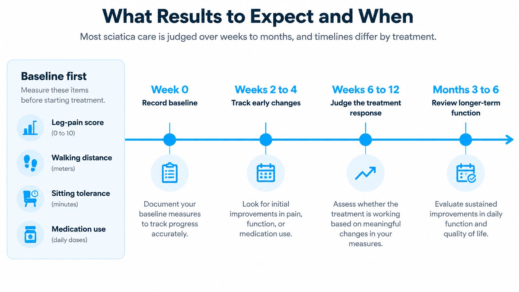
Start with a baseline before treatment: leg-pain intensity, walking distance, sitting tolerance, medication use, and a documented neurological examination. Choose activities you can repeat safely, such as the same walking route or a familiar seated task.
Timelines vary, and the checkpoints below are a monitoring framework, not recovery deadlines. Use the same measures each time so that a good day isn't mistaken for a sustained response.
The timeline illustrates that approach.
Separate monitoring dates from promised results
Clinic-reported expectations describe possible early improvement within 4 to 8 weeks, further progress over 3 to 6 months, and remote reviews around 3, 6, and 12 months; these are provider reports, not established trial outcomes, and should be checked against the clinic's sciatica treatment information.
Measurement schedule for sciatica treatment progress
| Timepoint | What to measure | Realistic expectation |
|---|---|---|
| Baseline, day 0 | Leg-pain score, walking distance, neurological exam, medication use | A documented starting point |
| 2-4 weeks | Night pain and sitting tolerance | Early change is possible, not required |
| 4-8 weeks | Walking distance, pain score, medication use | The clinic-reported early MSC window isn't guaranteed |
| 3 months | Function questionnaire and repeat neurological exam | Decide whether to continue, adjust, or switch |
| 6 months | Pain, function, return to work or activity | Assess durability; regenerative trial data remain limited |
At the planned review, compare progress with the goals you agreed on. If pain, walking, and function haven't improved meaningfully, revisit the diagnosis and strategy rather than adding treatment by default.
A useful result is something you can do more comfortably and consistently, not merely a more reassuring MRI description.
Long-term regenerative outcomes remain uncertain. Symptoms can also recur after a good response, so maintain an agreed rehabilitation and reassessment plan rather than assuming that improvement means degeneration has been reversed.
Dream Body Clinic offers a complimentary case review and MSC-based lower-back protocols for people considering regenerative care for degenerative disc disease and sciatica. Visit Dream Body Clinic with your imaging, treatment history, and symptom record, and request a written discussion of suitability, risks, evidence, and follow-up before deciding.