You've opened your MRI report, found the words medial meniscus tear, and started wondering whether you need surgery. Maybe your knee swelled after a twist on the court. Maybe it's been aching on stairs for months, without any obvious injury.
The next decision isn't surgery or no surgery. It's whether the tear matches your symptoms, whether a fragment is blocking movement, and whether the tissue can heal. Those distinctions also determine when rehabilitation or a stem cell consultation makes sense, and when an orthopedic surgeon should assess the knee first.
Table of Contents
- How a Medial Meniscus Tear First Shows Up
- What the Inner Knee Pad Actually Does
- When the MRI Tear Is Not the Pain
- Rehab Repair and Meniscectomy Choices
- How Stem Cells Guide Meniscus Healing
- Choose Knee Stem Cell Care or Surgery First
How a Medial Meniscus Tear First Shows Up
You plant your foot during a game and turn while your shoe stays put. The inside of your knee hurts, and swelling follows. An acute tear is one possibility, but an examination still needs to check the ligaments, cartilage, and other structures.
A different scene is just as familiar: you're past middle age, haven't fallen, and notice increasing discomfort when getting out of a low chair. Your MRI describes a degenerative tear. That finding calls for a different conversation from a displaced fragment that physically prevents straightening.
A medial meniscus tear is a split, fray, or detachment of the C-shaped pad on the inner side of the knee. Start by identifying the pattern rather than treating every tear as the same injury.
Medial meniscus tear patterns and first steps
| Tear pattern | Where it usually sits | Hallmark clue | First step |
|---|---|---|---|
| Acute traumatic tear | Posterior horn in most adult medial cases | Twist, swelling, inner joint-line pain | Exam plus MRI correlation |
| Degenerative tear | Inner knee, often with cartilage wear | Gradual ache after 40, no single injury | Activity change and rehabilitation first |
| Peripheral red-zone tear | Outer rim near the capsule | Tear in the vascular zone | Repair is often plausible |
| Inner white-zone tear | Central, poorly vascular rim | Fraying with little blood supply | Tissue-preserving plan; repair less likely |
| Posterior root tear | Back attachment on the tibia | Lost load sharing, sometimes extrusion | Prompt orthopedic assessment |
| Bucket-handle or displaced tear | Fragment flipped into the joint | Locking or inability to straighten | Same-week specialist care, sooner for a persistently locked knee |
These categories overlap. A traumatic tear can also be peripheral, and a degenerative tear can involve the root. The table helps you recognize the questions a clinician needs to answer, not diagnose the knee yourself.
Why the back of the meniscus gets attention
A review of meniscal injury and repair literature reports that adult medial injuries occur approximately twice as often as lateral injuries, with posterior tears accounting for approximately 93.1%–97.7% of adult medial-meniscus cases in the studies reviewed. The same review cites an annual incidence of about 66 meniscal tears per 100,000 people and approximately 850,000 meniscectomy procedures annually in the United States, illustrating the historical burden rather than how many people necessarily need surgery today.
Location matters when describing symptoms. Medial means the inner side, toward the other knee; lateral means the outer side. If your discomfort is mainly outside the joint, a guide to lateral knee pain can help you describe that distinction accurately.
Practical rule: A knee that won't fully straighten needs assessment, not repeated attempts to force it straight.
Major swelling after trauma or suspected root involvement also warrants prompt orthopedic attention. A stable knee without locking usually allows time for a planned examination and a rehabilitation discussion.
Keep the exact MRI wording, including terms such as posterior horn, radial, root, displaced, and extrusion. Those words describe more than the appearance of a split. They help explain whether this small pad can still share load between the thighbone and shinbone.
What the Inner Knee Pad Actually Does
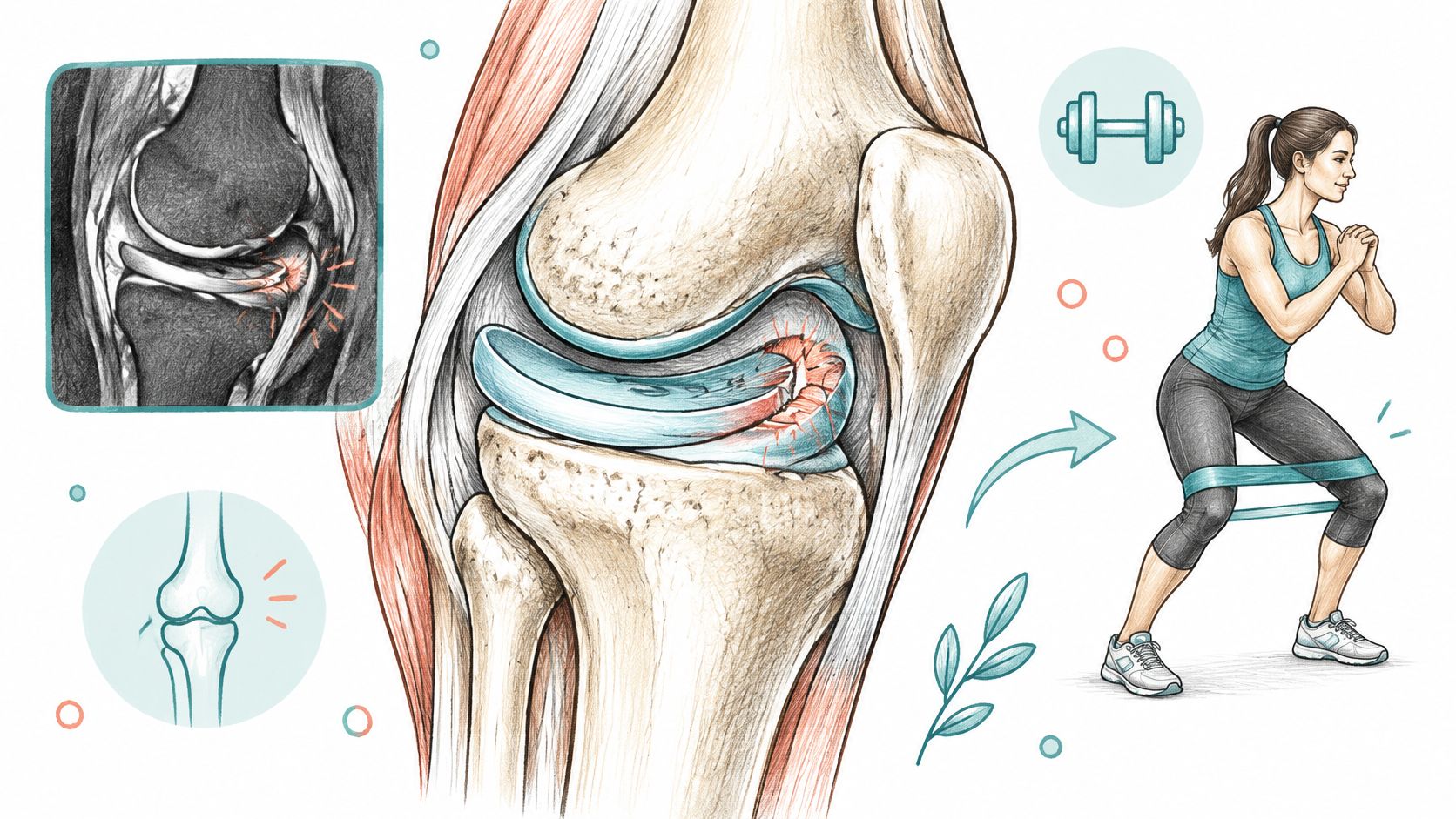
Think of the medial meniscus as a crescent-shaped gasket, not a loose pillow. It helps the rounded end of the femur, your thighbone, meet the flatter top of the tibia, your shinbone, across a broader contact area.
Say you step down from a curb. A functioning meniscus helps distribute that load instead of concentrating it on a smaller patch of joint cartilage. It also contributes to stability, which is why preserving useful tissue matters more than removing anything that looks irregular, as explained in AAOS patient guidance on meniscus tears.
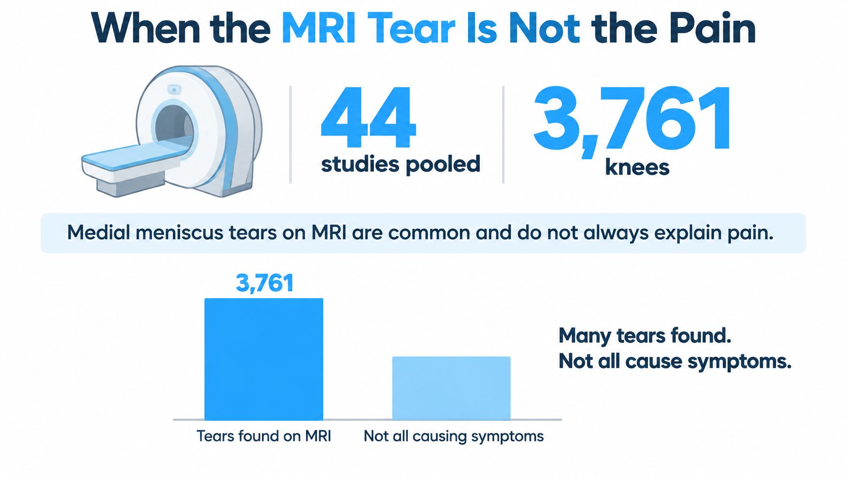
The illustration below introduces an important distinction: finding a structural change on an image isn't the same as identifying the cause of pain.
Blood supply changes the healing options
The meniscus doesn't receive blood evenly. Its outer edge, beside the joint capsule, has better access to circulation than its inner free edge.
Clinicians describe this map with color-based names:
- Red-red zone: The outer region has a blood supply that can support healing when the tear's shape and stability are suitable.
- Red-white zone: This transition region has less dependable vascular support, so repair decisions require closer attention to tissue quality.
- White-white zone: The inner region is relatively avascular. A central fray has less biological support for a conventional repair.
These aren't guarantees. A peripheral tear can still be too damaged to repair, while a surgeon may consider selected complex patterns using tissue-preserving techniques. Blood supply is one part of the decision, alongside whether the torn edges can be brought together and held securely.
A medial meniscus tear is both a structural injury and a biomechanical problem. The question is whether the remaining tissue can still do its job.
Why tear shape matters as much as size
The medial meniscus is relatively firmly attached, so it has less freedom to move during a planted-foot twist. Its posterior horn, the back portion, can be overloaded as the knee bends and rotates.
An acute sports split may occur in otherwise healthy tissue. A degenerative tear develops in a different environment, sometimes alongside cartilage thinning and changes in the quality of the meniscus itself. The same treatment won't necessarily suit both.
This video offers another way to visualize the knee and its treatment considerations.
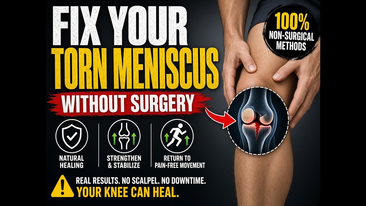
Before discussing treatment, circle location, zone, root, and displacement on your report, or ask the clinician to identify them. A small tear at an important attachment can matter more mechanically than a longer stable fray.
When the MRI Tear Is Not the Pain
A tear on MRI doesn't automatically explain a painful knee. A review discussing asymptomatic MRI findings summarizes a systematic review of 44 studies involving 3,761 asymptomatic, uninjured knees, with meniscal tears present in 10%, and a community study in which 61% of participants with a tear reported no knee pain, aching, or stiffness during the preceding month.
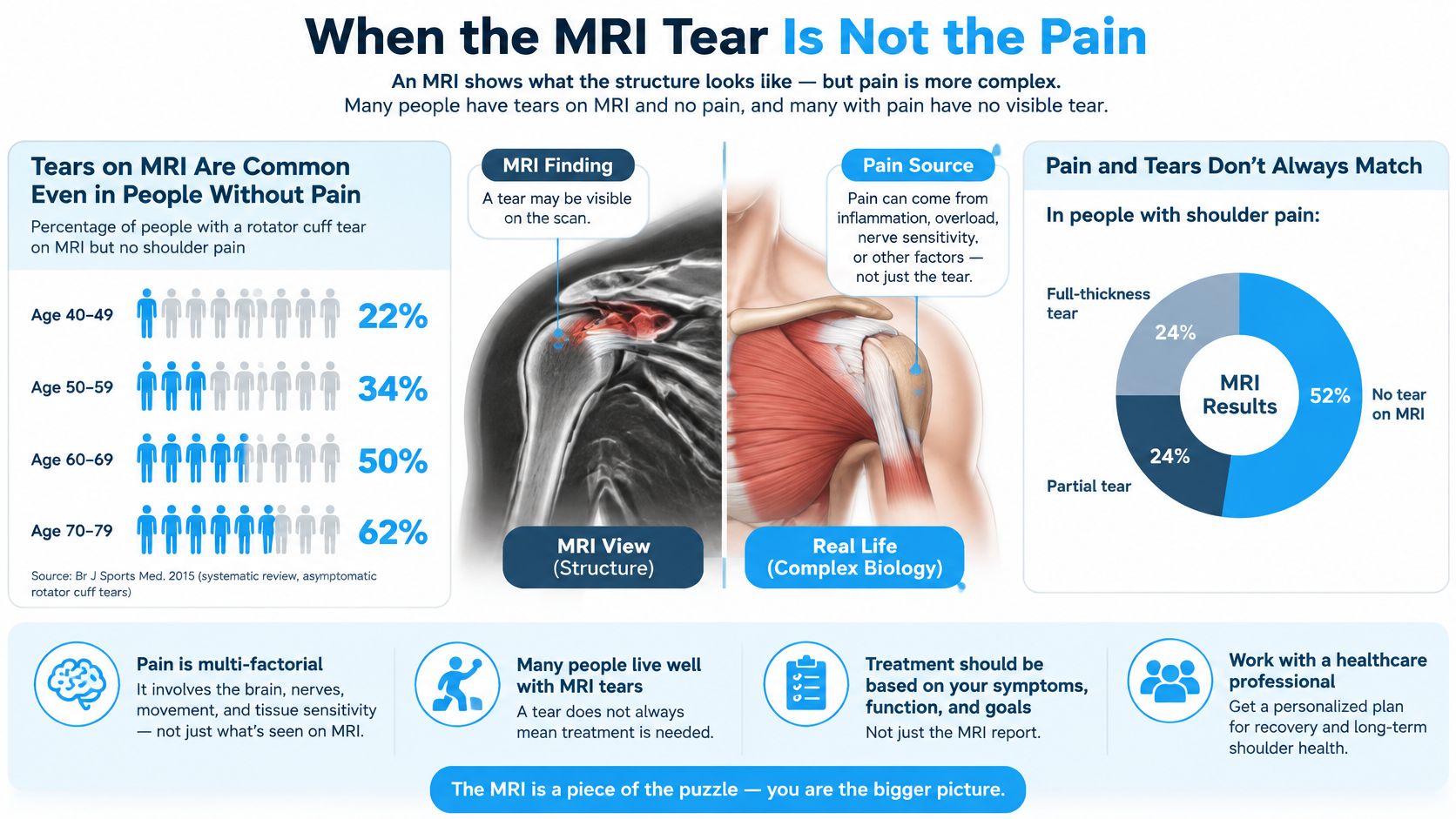
The figures below describe meniscal findings, not exclusively symptomatic medial tears. The community sample also wasn't restricted to people without symptoms.
Medial meniscus tear prevalence on MRI
| Group | Tear prevalence | 95% CI |
|---|---|---|
| Asymptomatic knees, 44 pooled studies (3,761 knees) | 10% | 7%–13% |
| Adults aged 40 and older in the asymptomatic review | 19% | 13%–26% |
| Adults younger than 40 in the asymptomatic review | 4% | 2%–7% |
| Community MRI, right knees, ages 50–90 (n=991) | 31% | 28%–34% |
| Participants with a tear reporting no knee pain, aching, or stiffness in the preceding month | 61% (180 of 297) | Not reported in the cited summary |
The confidence interval describes uncertainty around an estimate. It doesn't tell you whether your own tear hurts, and the final row describes the symptom status of people with tears, not tear prevalence in another population.
The supplied illustration uses shoulder imaging to reinforce the broader distinction between an imaging finding and symptoms; it doesn't provide additional knee statistics.
Match the image to the examination
MRI remains useful after an acute injury because it also evaluates cartilage, ligaments, and other soft tissues. A clinician combines those images with joint-line tenderness, the McMurray test, and the Thessaly test rather than relying on the report's label alone.
Prepare a symptom checklist:
- Location: Pain along the inner joint line.
- Rotation: Pain when twisting or pivoting.
- Movement: Catching, locking, or giving way.
- Activity response: Swelling that builds after activity.
- Loaded bending: Pain with deep squatting or stairs.
- Extension: A knee that stops fully straightening.
No single symptom settles the diagnosis. Greater agreement between the injury story, examination, and MRI location makes the tear a more convincing explanation.
Cartilage wear can also contribute to symptoms. Understanding knee arthritis pain and treatment helps separate pain from the surrounding joint from pain attributable to a meniscal lesion.
Findings that deserve prompt review
A persistently locked knee, inability to fully extend, major swelling after trauma, or report wording suggesting a root tear shouldn't be treated as routine aching. Root language deserves particular attention: a Mayo Clinic study of patients with subchondral insufficiency fracture of the knee found medial root or posterior-horn radial tears in 180 of 253 patients, or 71.1%.
That finding applies to a specific fracture population, not everyone with knee pain. It underscores why clinicians inspect the bone and attachments as well as the meniscus.
Bring your injury story, a short daily function diary, and the exact tear sentence from your MRI. Record what you can't do, what causes swelling, and whether the knee physically blocks or merely hurts during movement.
Rehab Repair and Meniscectomy Choices
For a stable degenerative tear, treatment usually starts by improving how the knee functions rather than changing its MRI appearance. That means adjusting painful activities, rebuilding strength, managing load, and controlling symptoms.
A clinical review of medial meniscal tear management supports conservative care first for degenerative tears and describes at least three months of nonoperative management before considering surgery, unless mechanical findings justify earlier review. An arthritis-associated tear without blocked motion doesn't automatically require an operation.
Care paths for a medial meniscus tear
| Care path | Best fit | What changes | Horizon or known limit |
|---|---|---|---|
| Rehabilitation and load management | Stable degenerative or non-locking tear | Strength, swelling, and daily function | A structured trial, unless locking, displacement, or root involvement changes urgency |
| Meniscal repair | Peripheral, reducible, viable tissue or a suitable root tear | Preserves tissue and shock absorption | Healing isn't assured; recovery requires protection and rehabilitation |
| Partial meniscectomy | Selected irreparable fragment causing a true mechanical block | Removes the obstructing piece, but also removes load-sharing tissue | Symptom relief is possible; later wear remains a concern |
| Mesenchymal stem cell injection | Selected partial or degenerative tear without a locked knee | Intended to influence the local healing environment through paracrine signaling | Clinical evidence is limited; assess symptoms, function, and appropriate follow-up imaging |
Make rehabilitation measurable
A useful program includes progressive quadriceps and hip strengthening, tolerable movement, and a plan for increasing walking, stairs, or work duties. Temporary bracing may help selected patients, but it doesn't reconnect a detached root or reposition a trapped fragment.
Don't judge the program only by how many appointments you attend. A randomized trial in adults with knee pain, osteoarthritis, and meniscal tears found that adding supervised standard physical therapy to home exercise didn't significantly improve three-month pain outcomes, with a KOOS pain difference of 2.5 points and a 98.3% confidence interval of −1.3 to 6.2.
That doesn't mean exercise is ineffective. It means a well-designed home program, adherence, and appropriate progression deserve attention, rather than assuming more clinic visits automatically produce a better result.
Before calling rehabilitation a failure: Check whether you followed a progressive plan and whether meaningful activities improved. Persistent pain deserves reassessment, not an automatic operation.
Preserve tissue when surgery is appropriate
Repair is attractive when the tear can be reduced, meaning the edges can be returned to a useful position, and the tissue can hold a repair. Keeping the meniscus preserves its mechanical role, but repair isn't guaranteed to last.
Evidence summarized alongside orthopedic guidance on acute meniscal pathology includes an overall repair failure rate of approximately 23.1% beyond five years, with approximately 24.2% after medial repair versus 20.2% after lateral repair. Suitable root repairs may improve pain and function without guaranteeing prevention of later X-ray arthritis.
Partial meniscectomy has a narrower purpose: removing selected irreparable tissue when its mechanical effects justify the loss. It isn't a default response to an MRI finding, and vague clicking alone isn't the same as a true mechanical block.
A locked or root-pattern knee should reach an orthopedic surgeon before anyone proposes injections as a substitute for correcting the mechanical problem.
How Stem Cells Guide Meniscus Healing
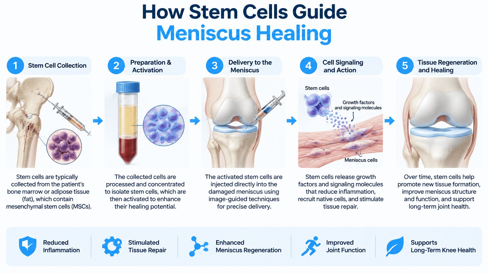
Mesenchymal stem cells, or MSCs, are best understood here as signaling cells, not replacement meniscus material. The treatment rationale isn't that they differentiate into other cell types. It's the paracrine effect, meaning they release specialized signals that guide nearby cells and the local healing environment.
Those signals include cytokines, chemokines, and extracellular vesicles. A review of MSC signaling mechanisms describes effects on cell survival, migration, inflammation, and extracellular-matrix activity, while noting limited cell survival and engraftment at injured sites.
What a local treatment is intended to do
The extracellular matrix is the supportive material surrounding cells. Influencing its maintenance and repair is different from placing a ready-made patch into a tear, and it can't be assumed to overcome a displaced fragment or detached attachment.
At our clinic, we place cells locally at the injury site rather than relying on IV delivery for cartilage. Our rationale is that cartilage receives little blood flow, limiting delivery through circulation; local placement targets the affected area, where inflammatory signaling and fibrosis, or scar tissue, are treatment targets. That rationale shouldn't be mistaken for proof that every injected tear will regenerate.
This diagram outlines the assessment and treatment process.
Favorable findings and what they establish
A systematic review of stem-cell applications for meniscal tears identified 18 studies, including 13 preclinical and five clinical studies, with clinical follow-up of three to 24 months; all clinical studies reported favorable laboratory, clinical, or imaging findings, and bone-marrow-derived MSC injection was the most frequently studied approach. The authors still called for larger randomized trials and longer follow-up.
Small clinical reports provide encouraging but narrower evidence. A review of synovial MSC transplantation describes five men with complex degenerative medial tears, mean age 48.2 years, whose pain, daily-activity, and sports scores improved two years after repair plus transplantation, and another series involving six patients, five men and one woman, mean age 54 years, treated with repair plus synovial MSC transplantation.
Because surgery and cells were combined, those reports don't establish that an injection alone would produce the same result. Cell source and delivery method also differ between protocols.
A systematic review of meniscus regeneration describes a human report using 16 million adipose-derived cells with platelet-rich plasma and hyaluronic acid for a grade-II tear, with reduced pain but minimal tissue regeneration at three months. Pain improvement and structural restoration are separate outcomes.
Keep individual cases in perspective
Our clinic reports that Josh's baseline MRI showed a partially torn ACL and multiple meniscus tears, and that a follow-up MRI nine months after treatment showed healing. This is an individual clinic-reported case, not an independently verified controlled comparison or a guarantee for another patient.
For selected partial or degenerative tears, a consultation can examine whether this approach fits the knee's actual structure and symptoms. A locked knee, displaced flap, or root tear still needs surgical review first.
Choose Knee Stem Cell Care or Surgery First
Choose prompt orthopedic assessment when the knee stays locked, won't fully extend, swells heavily after trauma, or the MRI describes a displaced bucket-handle or posterior root tear. The immediate priority is determining whether the joint has a mechanical problem that needs correction.
A regenerative consultation is more appropriate for a selected partial or degenerative tear without mechanical locking, particularly when a structured rehabilitation program hasn't restored useful function. You can also seek an opinion before accepting surgery, provided that consultation doesn't delay necessary treatment.
What our knee assessment includes
Dream Body Clinic in Bucerias, Nayarit, Mexico, offers a knee stem cell treatment for selected partial meniscus injuries, cartilage wear, and partial ACL, PCL, or MCL injuries. Our protocol uses 50 million mesenchymal stem cells, and the package costs $3,500 USD per knee, including an MRI and blood panel.
We deliver care in Mexico within a COFEPRIS-regulated framework. COFEPRIS is Mexico's federal health regulator; regulatory oversight doesn't by itself establish that a treatment will repair a particular tear.
Visits typically take one to three hours, with remote check-ins around three, six, and 12 months. Before making travel arrangements, review our information on stem cell therapy safety for orthopedic injuries and discuss the proposed approach with your own doctor.
Make the decision around function
A useful treatment goal is specific: walking without recurrent swelling, returning to necessary work tasks, or managing stairs comfortably. Improvement should be assessed against those goals, not only against a scan.
Outcomes vary, the clinical evidence remains limited, and no result is guaranteed. Send your existing MRI images and report for a complimentary case review so we can assess whether a regenerative consultation is appropriate or whether orthopedic care should come first.
Dream Body Clinic offers MRI-informed assessment and targeted knee stem cell care for selected partial meniscus injuries. Visit Dream Body Clinic to request a complimentary review of your MRI and symptoms, and discuss the recommendation with your treating doctor before deciding on treatment or travel.