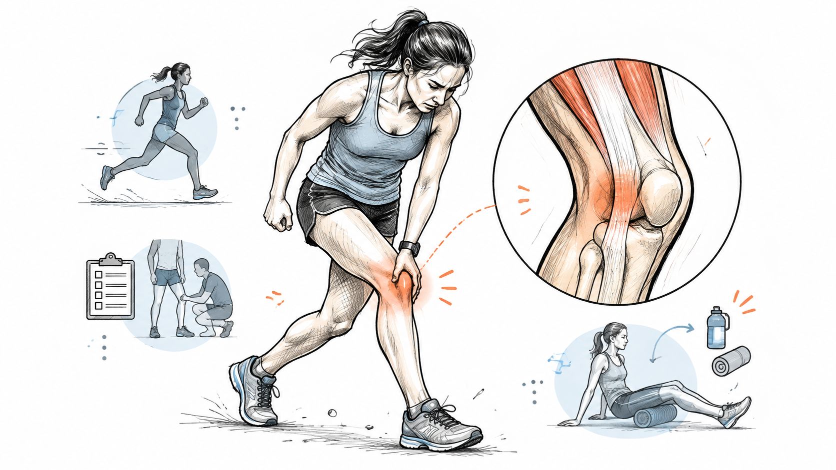
Two runners press a finger into the same square centimetre of skin, two or three centimetres above the outer knee joint, and describe the same sharp pain. Do they have the same injury? Often they don't. One has an irritated iliotibial band. The other has a lateral meniscus tear, and the spot that hurts is where the joint line sends its ache.
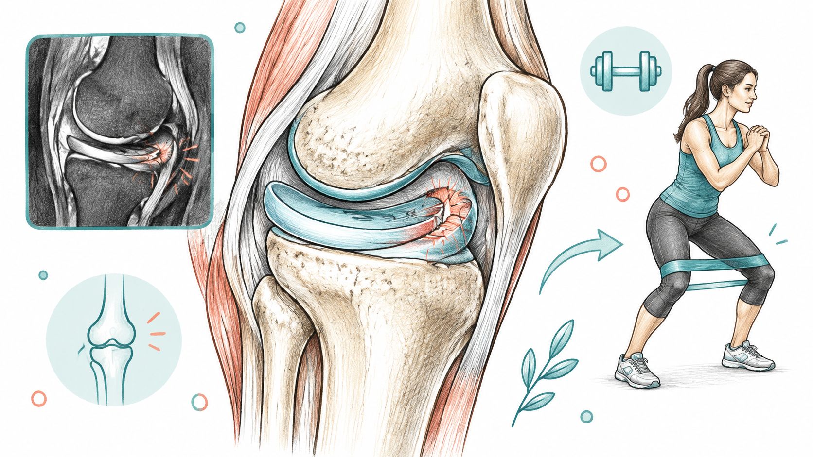
The outer knee is a crowded place. The iliotibial band runs down to Gerdy's tubercle on the outer shin, the lateral collateral ligament braces the joint, the lateral meniscus cushions it, and the popliteus and biceps femoris tendons attach around the back corner. Pain can also arrive from the hip or the lumbar spine with nothing wrong at the knee itself.
What separates these conditions isn't the address of the pain. It's the pattern: when it started, what distance or movement brings it on, whether the knee swells, catches or gives way. Read that pattern and you'll know what to ask for.
Table of Contents
- Lateral Knee Pain Causes and How to Read Your Pattern
- Iliotibial Band Syndrome in Runners and Cyclists
- Trauma, Locking and Warning Signs to Act On
- What an MRI Can and Cannot Tell You
- Treatment Options Matched to Each Diagnosis
- Get the Right Assessment Before You Choose Treatment
Lateral Knee Pain Causes and How to Read Your Pattern
Lateral knee pain names a place on the leg. It does not name a cause. In runners the most frequent cause is iliotibial band syndrome, an overuse problem in the tissue along the outer thigh, and a clinical review of lateral knee injuries identifies it as the most common injury on the lateral side of the knee in runners.
The same review ties the condition to distance running, cycling and military training rather than a sedentary life. Repetitive bending under load is the shared ingredient across those sports. The catch is that active people also tear menisci, sprain ligaments, wear down joint cartilage and feel pain referred from the hip or spine. The address of the pain alone will never separate those causes.
The table below does the separating. Read across each row and notice how differently the conditions behave.
| Cause | Typical pain pattern | Key clue | First step |
|---|---|---|---|
| Iliotibial band syndrome | Sharp or burning outer knee pain that starts after a set distance | Predictable onset, worse on downhill or cambered surfaces | Reduce load and progress hip and knee strengthening |
| Lateral meniscus tear | Joint-line pain with clicking, catching or locking | Often follows a twist or deep squat | Clinical examination, then MRI if mechanical symptoms persist |
| Lateral compartment osteoarthritis | Ache after activity and morning stiffness | Worse on stairs and on rising from a chair | Weight-bearing X-rays and supervised rehabilitation |
| Lateral collateral ligament injury | Outer knee pain with instability or giving way | Usually follows a direct blow or twist | Examination of the collateral ligaments and imaging |
| Popliteus or biceps femoris tendinopathy | Pain at the back of the outer knee, worse on descending | Tenderness at the tendon attachment | Load reduction and graded loading |
| Referred hip or lumbar pain | Lateral thigh and knee ache with a normal knee examination | Pain changes with hip or back movement | Hip and spine examination |
| Stress fracture | Localized bone pain that worsens with impact | Pain that progresses during running | Imaging for fracture and rest from impact |
| Urgent or systemic causes | Pain at rest, night pain, fever, warm and swollen knee | Systemic signs or inability to bear weight | Same-day medical assessment |
The first row is the common running answer and the one many people arrive with. The meniscus and ligament rows follow a twist, a deep squat or a blow, and they bring swelling, clicking or a knee that buckles. The urgent row exists because fever, rest pain and a knee that can't take weight don't belong in a self-management plan. Even a scan needs this context, since a finding on imaging only matters if it matches the pattern in front of the clinician.
A four-question check to narrow the list
1. Where exactly is the sore spot? Tenderness sitting just above the outer joint line, over the bony point on the side of the thigh, points toward the band. Pain right on the joint line points toward the meniscus. A knee that gives way suggests the lateral collateral ligament, and pain at the back of the outer knee that worsens on descents fits the popliteus or biceps femoris tendon.
2. When does it start? Pain that arrives at the same distance on every run, then earlier each week, is the signature of an overuse problem. Pain that began after a twist or a deep squat points to a structural injury, even if it felt mild at the time.
3. What loads it? Morning stiffness that eases with movement, aching on stairs and pain on rising from a chair fit osteoarthritis better than tendon trouble. Arthritis is graded in stages, and the guide to knee arthritis stages shows what each grade means for treatment.
4. Does it spread? Ache that runs up into the thigh or down toward the buttock, or a knee that hurts without any knee tenderness, can be referred from the hip or lumbar spine. Examining the hip and back matters far more here than stretching the knee.
The clue you can gather yourself: the pattern of onset and triggers. It's free, it's reliable, and it changes the list of likely causes before anyone touches your knee.
What to record before you're assessed
Write down four things: the date the pain started, your weekly running distance before it began, any change in training across the two weeks before onset (mileage, hills, speed, surface, shoes), and whether the knee swelled, locked or gave way. A note like this turns a vague complaint into a timeline the clinician can test against each cause in the table.
The check sorts patterns. It can't replace a hands-on examination, and it can't clear the conditions in the last row.
Iliotibial Band Syndrome in Runners and Cyclists
The iliotibial band is a thick strip of fascia, the connective tissue that continues down from the tensor fascia lata, gluteus medius and gluteus maximus and inserts mainly at Gerdy's tubercle on the outer shin. It behaves like a tensioned strap: the hip muscles set the tension, and the outer knee feels the pressure when that strap is repeatedly loaded.
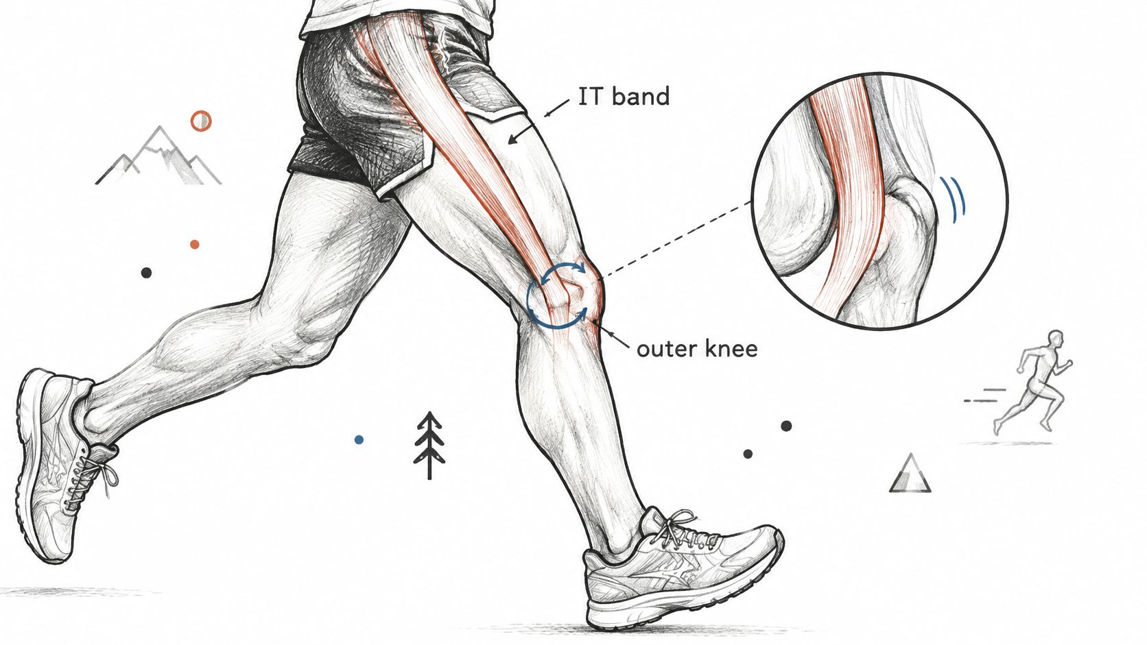
Pain usually sits 2 to 3 centimetres above the lateral tibiofemoral joint line, near the lateral femoral epicondyle. What reproduces it is movement: as the knee bends and straightens, tissue compresses and shifts across that bony point, and clinicians find the tender spot with the knee near 30 degrees of flexion, close to where the knee sits at foot strike in running, as described in a clinical review of iliotibial band syndrome. That timing explains why the pain often appears mid-run rather than at the start, and why it builds over weeks instead of arriving in one moment. Outer location narrows the list, but the repeating trigger is what earns the diagnosis.
The condition accounts for up to about 12% of running-related injuries and shows up in cycling and military training too, where the same repetitive bending loads the same spot. Sport changes the size of the problem: one review cited 24% occurrence among road cyclists, according to a review of iliotibial band syndrome frequency across sports.
Turning each trigger into a change
- A jump in weekly mileage. Hold volume flat for two weeks, then add distance back in smaller steps than before.
- Downhill running and hill work. Walk the steep descents for now and reintroduce them after two pain-free weeks.
- A longer stride. Shorten it slightly and let cadence rise, so the knee spends less time near the compressed position.
- Cambered roads. Run the flat side of the road or switch to a path, because a constant tilt loads one outer knee harder.
- Higher intensity. Keep one hard session a week instead of stacking several.
What hip strength does and doesn't explain
Hip weakness gets blamed for almost every outer knee. The evidence is narrower than the reputation: studies looking for a single hip-strength cause have produced mixed results, and the link between weakness, pelvic control and symptoms is less consistent than runners are often told.
Test the hip rather than assuming it. If abduction strength is genuinely low on one side, strengthening earns its place. If it isn't, adding more hip work won't fix a load problem.
The practical order is unglamorous: cut the provoking load for a week or two, avoid abrupt jumps in mileage or hills, look at foot posture and running pattern, then build hip and lower-limb strength in graded steps and maintain it.
Trauma, Locking and Warning Signs to Act On
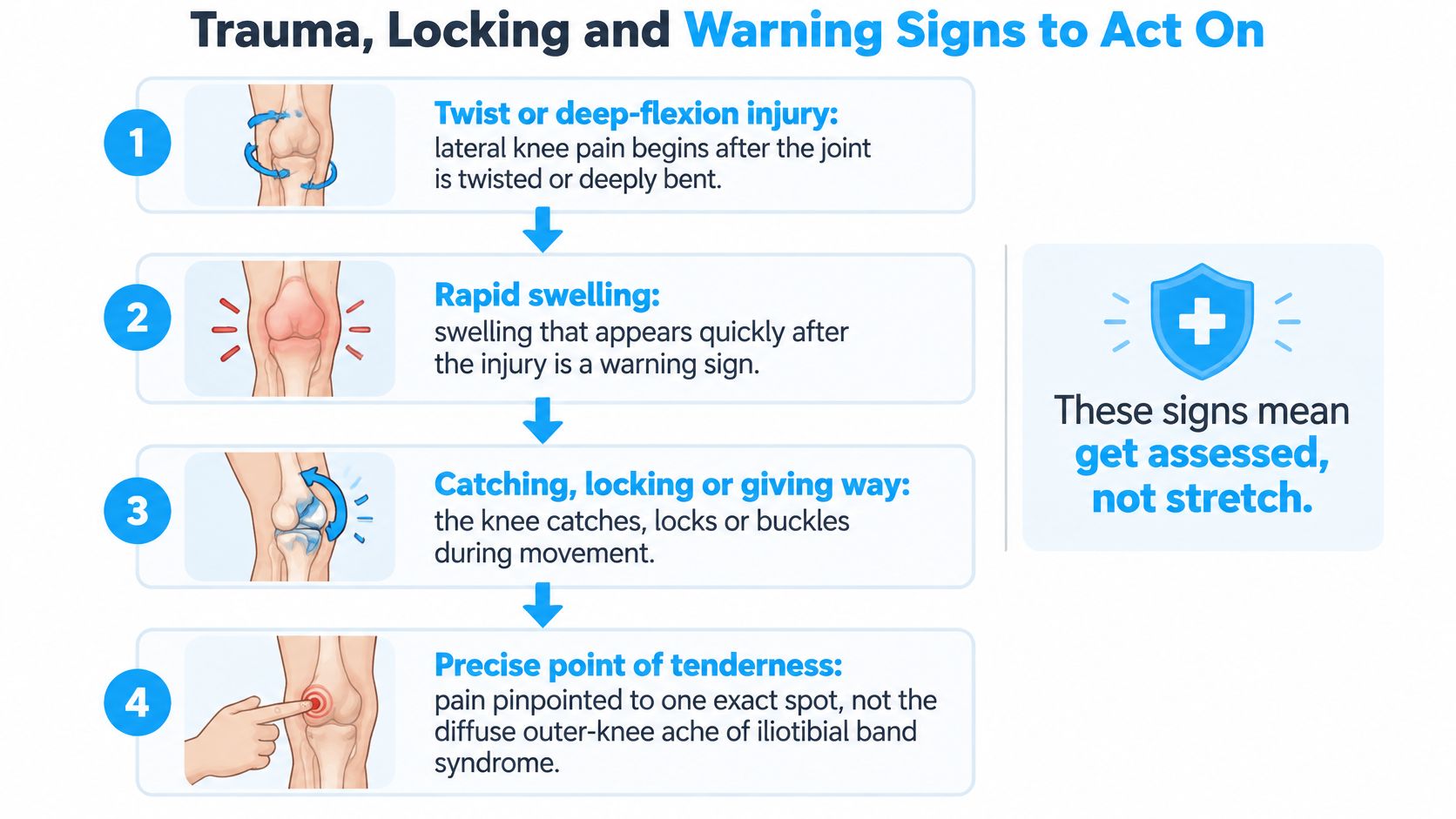
Outer knee pain that arrives after a twist is a different animal from outer knee pain that arrives after a long run. When lateral pain follows trauma, particularly with rapid swelling, catching, locking, instability or tenderness pinpointed to one spot on the joint line, the working diagnosis should be a lateral meniscus or ligament injury until an examination says otherwise. Labelling it a tight band delays the assessment that matters, as the American Academy of Family Physicians review of knee pain evaluation sets out.
The red flags that mean same-day care
- Fever or a hot, swollen knee. Infection and inflammatory disease both need urgent assessment.
- Inability to bear weight after an injury points to fracture or major structural damage.
- Visible deformity of the knee or calf.
- Calf swelling and pain, which raises the question of a clot.
- A knee that locks and won't straighten fully.
What the examination looks for
Clinicians test the meniscus with the Thessaly maneuver, generally preferred over McMurray testing for suspected meniscal injury, though acute pain or instability can limit either one. Effusion that appears within hours of a twist raises suspicion of internal derangement far more than swelling that creeps in over days.
The differential stays wide: hip disease, lumbar referred pain, patellofemoral disorders, gout, infection, osteoarthritis, ligament injury and iliotibial band syndrome all produce pain around the outer knee. A meniscus tear can also cause catching with forceful bending or straightening.
A triage order that works
- Screen for red flags first. Fever, marked warmth, inability to bear weight, deformity, calf swelling or a locked knee means urgent care, not a stretching program.
- Characterize onset and mechanical symptoms. A twist, a deep squat, a pop, a catch, a buckle. Each word changes the list.
- Examine in a fixed order. Joint line, collateral ligaments, patellofemoral region, hip, lumbar spine.
A locked, unstable or non-weight-bearing knee gets seen promptly. A meniscus that keeps catching deserves assessment of a complex meniscus tear rather than another month of foam rolling.
What an MRI Can and Cannot Tell You
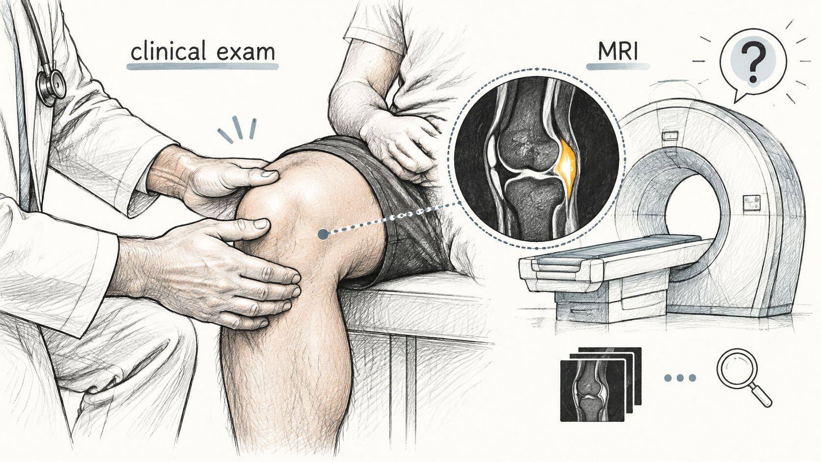
The question that follows an MRI report is almost always the same: does this finding explain my pain, and will an operation fix it? The honest answer starts with what each scan can do. Iliotibial band syndrome is usually diagnosed clinically. MRI can support the diagnosis, but routine radiographs don't confirm it, though they do reveal competing causes such as osteoarthritis or a stress fracture, as a review of imaging in lateral knee pain explains.
Which scan answers which question
Weight-bearing X-rays when fracture or osteoarthritis is suspected. They show joint space, which is where lateral-compartment wear becomes visible.
MRI when mechanical symptoms persist, when a traumatic effusion needs explaining, or when suspected internal derangement would change management. It defines meniscal and ligament injury better than any other study.
Ultrasound, in trained hands, for tendons, bursae and superficial structures.
An abnormal scan is not automatically the pain generator
This is the part patients rarely hear before a procedure gets booked. A lateral meniscus tear on MRI and a signal change in the band both appear in knees that hurt elsewhere, and neither explains pain on its own. Joint-line pain, catching and locking carry more weight than an isolated scan finding. Treatment decisions belong to the pain pattern, the functional limitation, the examination and the mechanical symptoms, not to the image alone.
Ask three questions before agreeing to anything invasive:
- Which finding on this scan matches the pain I describe?
- What would you recommend if the scan were normal?
- What does a supervised rehabilitation period look like, and when do we review it?
A scan describes tissue. It doesn't describe your pain. The clinician's job is to connect the two, and sometimes the honest conclusion is that they don't connect.
Non-operative care deserves a defined trial: a loading and strengthening program with a review point set in advance, not an open-ended wait. Escalate for a surgical opinion if swelling persists, the knee locks, instability interferes with daily life, or night and rest pain continue. Rest pain without an obvious injury isn't a routine tight-band story and deserves assessment.
Treatment Options Matched to Each Diagnosis
Treatment follows the diagnosis. An injection that helps arthritic cartilage does nothing for a tendon compressing against bone, and a strengthening program that settles a band problem won't stabilize a torn ligament. The table sets first-line care and the regenerative option worth discussing for each one.
| Diagnosis | First-line care | Regenerative option to discuss | Evidence note |
|---|---|---|---|
| Iliotibial band syndrome | Load reduction, graded return to running, hip and knee strengthening | Not a stem cell indication based on current evidence | Clinical diagnosis with supervised rehabilitation |
| Lateral meniscus tear | Activity change, targeted rehabilitation, orthopedic review for locking | Discuss with the treating team if mechanical symptoms persist | Judged by matching symptoms to MRI findings |
| Lateral compartment osteoarthritis | Strengthening, weight management, pain control | Intra-articular MSC injection, discussed once imaging confirms arthritis | Meta-analysis of 502 patients showed WOMAC and pain gains |
| Lateral collateral ligament injury | Bracing, graded rehabilitation, stability testing | Specialist review if instability persists | Based on injury grade and examination |
| Popliteus or biceps femoris tendinopathy | Load management, isometric then progressive loading | Not an established stem cell indication | Rehabilitation-based care with limited trial data |
| Referred hip or lumbar pain | Treat the source identified on examination | Hip or spine treatment chosen by a specialist | Set by the confirmed source |
Stem cells work by signalling, not by becoming new tissue
Mesenchymal stem cells don't turn into cartilage, meniscus, ligament or nerve cells. Their action is paracrine: they release specialized signalling molecules, extracellular vesicles and immunomodulatory factors that influence inflammation, pain signalling and the local repair environment. The tissue isn't rebuilt cell by cell. The environment around it changes.
The strongest evidence sits with knee osteoarthritis. A 2025 systematic review and meta-analysis of mesenchymal stem cell injections for knee osteoarthritis pooled eight randomized controlled trials with 502 patients and found intra-articular MSC injection improved WOMAC function and symptom scores by 7.44 points at six months (95% CI, 1.45 to 13.42; P = 0.01) and 10.31 points at twelve months (95% CI, 0.96 to 19.67; P = 0.03) versus controls, with gains in visual analogue pain scores and KOOS outcomes and no meaningful increase in adverse events.
Scope matters. That evidence covers knees with degenerative joint disease, including lateral-compartment wear that shows up as outer knee pain. It doesn't cover isolated iliotibial band irritation, and the trials are small, so individual results vary.
The distinction to hold onto: a treatment can be reasonable for one diagnosis and irrelevant for another that hurts in exactly the same place.
Where a clinic fits into the decision
Once imaging confirms arthritis and a clinician agrees non-surgical options are appropriate, an intra-articular injection becomes a legitimate conversation. Dream Body Clinic's Stem Cell Knee Treatment places 50 million stem cells into the knee joint in a single injection that typically takes a few minutes. Many patients notice early improvement within 2 to 4 weeks, with fuller results developing over 3 to 6 months, and results vary. The procedure is low-risk with minimal chances of complications or infections.
Before treatment, the medical team examines the knee joint and discusses your history and expectations. The clinic lists arthritis and partial meniscus or ligament tears among the reasons people come; the published evidence supports the arthritis case, so a partial tear is a discussion point rather than a proven indication. Treatment happens in Mexico under COFEPRIS oversight, the country's equivalent of the FDA.
For a fuller picture of the non-surgical side, the knee arthritis pain treatment overview covers what else belongs in a plan.
Get the Right Assessment Before You Choose Treatment
Four steps, in this order.
- Log the pattern for two weeks. Record each episode with its date, the activity or distance that brought it on, any swelling, any catching or locking, and whether night pain woke you.
- Book a clinical assessment with a sports medicine or orthopedic clinician. Bring the log and every image you've had, plus the written report, not just the disc.
- Ask which diagnosis was reached and why. Then match it to the treatment table. If someone recommends an injection without naming a diagnosis, get the diagnosis first.
- If imaging confirms knee osteoarthritis and the clinician agrees non-surgical options are appropriate, you can ask about a consultation at Dream Body Clinic in Bucerias, Nayarit, where follow-up appointments can be scheduled to monitor progress and address concerns.
A consultation is a review, not a promise, and results vary. Before any of it: a locked knee, an inability to bear weight, fever or spreading swelling means seeking care now, ahead of every elective decision.
If your outer knee has hurt for months and the scan you were handed never matched the pain you feel, Dream Body Clinic starts where the imaging report stops: a clinical review of your knee, your training history and your scans, before anyone discusses treatment. Its stem cell knee treatment delivers 50 million mesenchymal stem cells into the joint in a single injection, with the medical team working through which findings on your images explain your pain and which ones don't. Visit Dream Body Clinic to start a case review.