You open your MRI report and find the words meniscus complex tear. Your knee may hurt on stairs, catch when you turn, or swell after a walk, but the report sounds more alarming than the symptoms you can describe. It's natural to wonder whether complex means severe, whether surgery is unavoidable, and whether an injection could help.
The useful answer starts with the knee, not the label. A clinician needs to connect the tear's shape with its blood supply, stability, surrounding cartilage, and your symptoms before recommending treatment.
Table of Contents
- What Makes a Meniscus Complex Tear Different
- Symptoms and Daily Impact of a Complex Tear
- How Doctors Confirm a Complex Tear
- Repair, Meniscectomy or Nonoperative Care Compared
- How Regenerative Injections Work on a Meniscus Tear
- Recovery Timeline After Repair or Regenerative Care
- Choose Your Next Step for a Complex Tear
What Makes a Meniscus Complex Tear Different
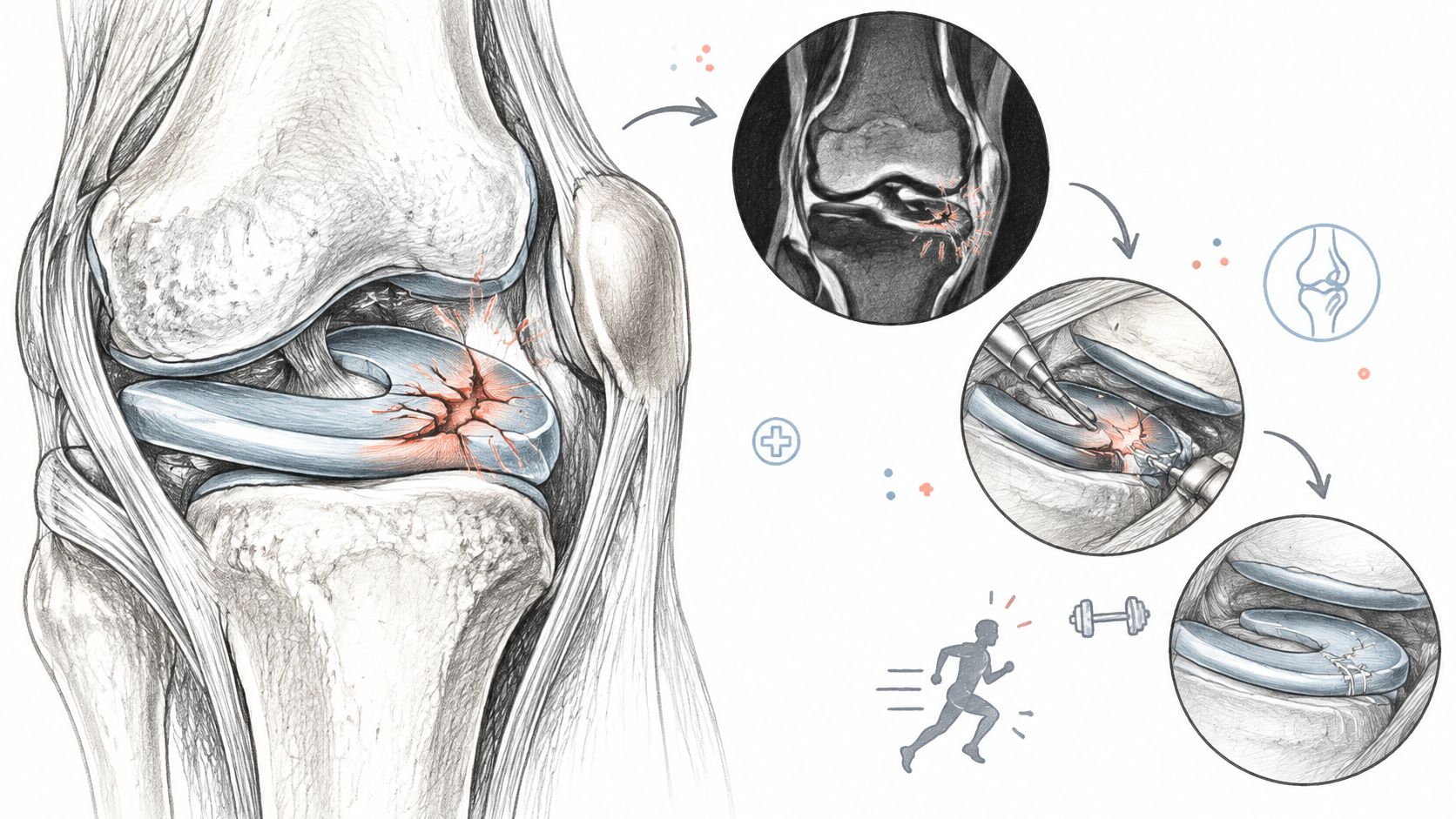
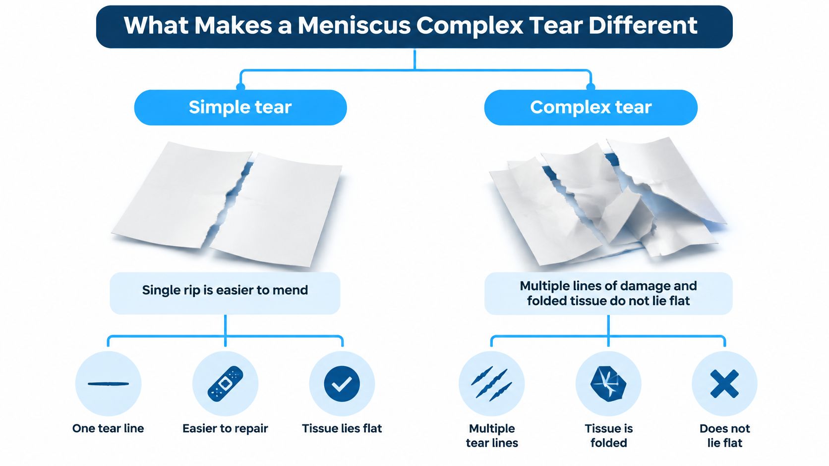
A complex tear is easier to understand if you separate the label from the tissue. The word describes geometry: the tear runs in more than one direction. It often combines horizontal, radial, longitudinal, or flap components, so the torn edges do not line up like a single clean rip in a sheet of paper.
A complex meniscus tear is not automatically a surgical tear. The MRI label tells you what the tear looks like. It does not tell you how well that piece of meniscus still carries load, whether a fragment shifts when the knee bends, how much blood supply reaches the torn edge, or whether your symptoms match the image. Those details shape the treatment decision more than the word "complex" does, and your age and symptoms are part of that picture.
Why the shape matters
The meniscus distributes pressure between the thighbone and shinbone. Its curved fibers carry tension around the knee, a function called hoop stress. If those fibers are cut, the meniscus can lose that tension and move outward, or extrude, so it spreads weight less effectively. The effect is similar to a cut hoop on a wooden barrel: the staves no longer stay pressed together as they should.
A flap can also move differently from the rest of the meniscus. That distinction matters because a stable split and a displaced fragment can require very different care, even when both reports use the same word. A fresh tear in a younger knee with a clear injury is also a different problem from a degenerative complex tear in an older knee, even when the MRI line reads the same.
Runners can find useful movement context in Swift Running's a runner's guide to knee mechanics. It is background education, not a way to judge tear stability yourself.
How common is this MRI pattern?
A 2021 epidemiological study of symptomatic knees classified 214 of 1,368 MRI-confirmed meniscal tears as complex, reported as 15.5%, with the selected pattern counts summarized below.
Meniscal tear patterns on MRI in symptomatic knees
| Tear pattern | Cases | Share of tears |
|---|---|---|
| Horizontal, oblique or undersurface | 577 | 42% |
| Complex | 214 | 15.5% |
| Radial | 187 | 13.6% |
| Bucket-handle | 88 | 6.4% |
| Other patterns, not itemized | 302 | 22.1% |
These findings describe people with symptomatic knees undergoing MRI, not all adults. The classification describes appearance, not an automatic treatment decision. A complex tear can be stable or unstable, painful or quiet, repairable or not, and the MRI alone does not settle which. Regenerative injections follow the same rule: they work by signaling the knee's own repair response, not by turning into new meniscus tissue.
Symptoms and Daily Impact of a Complex Tear
Say you can walk comfortably in a straight line, but turning away from the kitchen counter causes sharp pain along the inside of your knee. Later, the knee feels puffy, and standing after a long meeting takes a few careful steps. That pattern is more informative than telling a clinician that your knee hurts.
Meniscal symptoms can include joint-line pain, swelling after activity, stiffness, and discomfort with twisting or deep bending. Some people notice a clear injury; others develop symptoms gradually as tissue degenerates.
Catching isn't the same as locking
A click doesn't, by itself, prove that torn tissue is trapped. Knees can click for several reasons, and a giving-way sensation can come from pain, muscle inhibition, or ligament instability.
True locking means the knee physically won't move through its usual range, particularly when you try to straighten it. Don't repeatedly force it straight or use twisting exercises to free it.
Daily limitations often reveal the problem more clearly than pain intensity:
- Stairs: Descending may feel uncertain or uncomfortable.
- Deep bending: Kneeling, squatting, and rising from a low chair may provoke pain.
- Uneven ground: You may shorten your stride or avoid turning on the affected leg.
- Work and sport: Tasks involving crouching, carrying, or pivoting may become difficult.
Compression can improve comfort for some people, but it doesn't reposition displaced tissue. NZ Supps' guide to choosing between sleeves and wraps can help explain support options, provided you don't treat support as permission to push through worsening symptoms.
When to seek same-day care
Arrange urgent assessment for:
- A locked knee: You can't straighten it.
- Inability to bear weight: Standing or walking is no longer possible.
- Rapid swelling after injury: Especially with marked pain or instability.
- A hot, red knee with fever: This needs assessment for causes beyond a meniscal tear.
- A cold or numb foot: Seek emergency care, particularly with color changes.
For nonurgent symptoms, keep a brief movement log. Record what you did, where the pain occurred, and whether swelling followed. That gives the clinician a practical way to compare your experience with the MRI.
How Doctors Confirm a Complex Tear
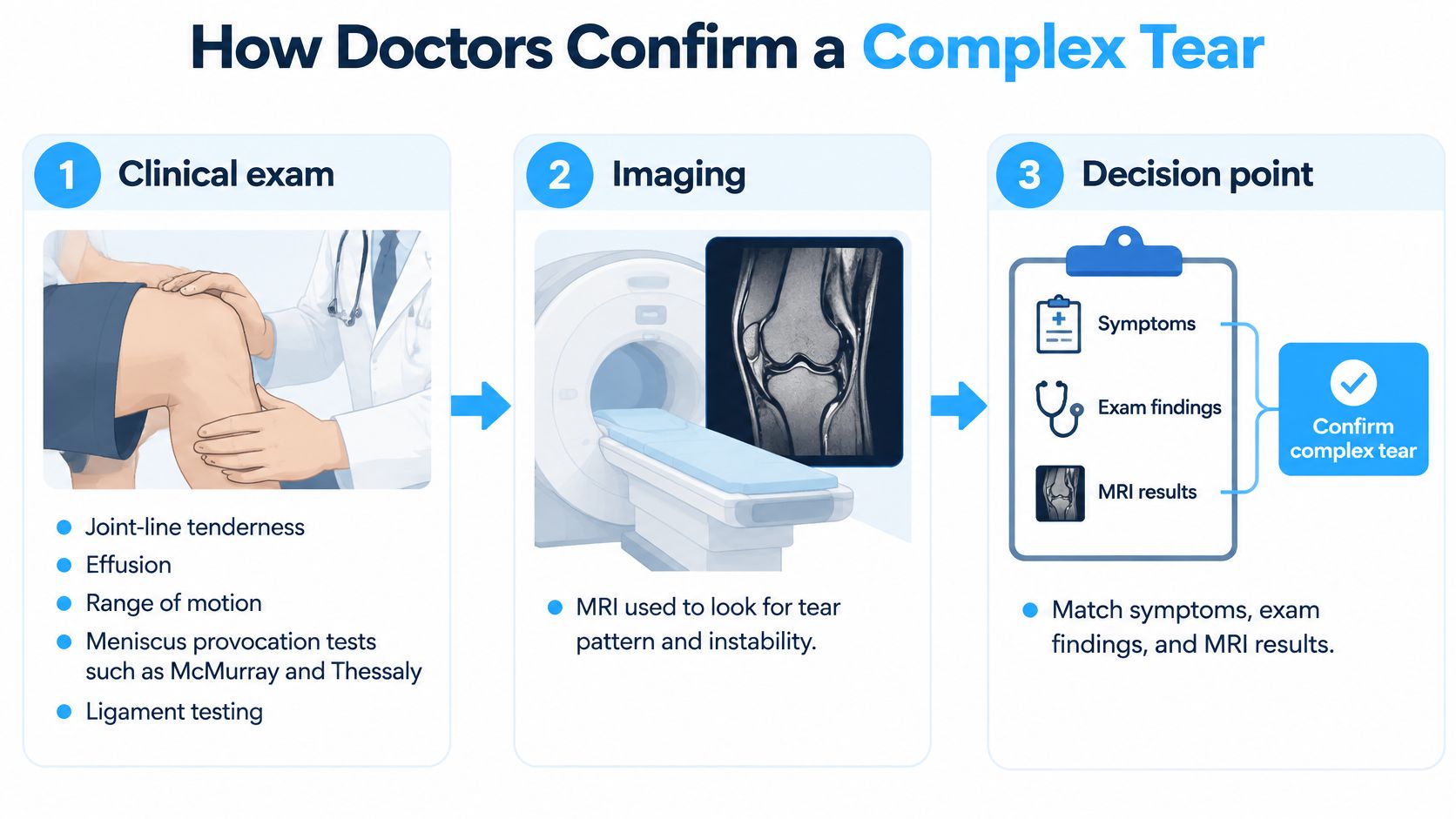
The examination begins with your story: whether you twisted the knee, how quickly swelling appeared, and what movements you can no longer perform. A clinician then checks joint-line tenderness, effusion, range of motion, and ligament stability.
Tests such as McMurray and Thessaly may help reproduce meniscal symptoms. They're interpreted together, rather than used as a stand-alone diagnosis, and they aren't exercises to try forcefully at home.
What MRI can establish
MRI helps map the tear's extent, displaced tissue, extrusion, cartilage damage, and associated ACL injury. A clinical review of meniscal tears reports the following approximate diagnostic performance and notes that extensive tears greater than 1 cm, particularly in younger patients or with acute ACL injury, commonly warrant surgical evaluation.
| Tear location | Sensitivity | Specificity |
|---|---|---|
| Medial meniscus, inner side | Approximately 89% | Approximately 88% |
| Lateral meniscus, outer side | Approximately 78% | Approximately 95% |
Sensitivity describes how well a test detects tears that are present. Specificity describes how well it identifies people without a tear. These figures mean MRI is useful, but it can miss injuries, particularly lateral tears, and can't settle every diagnosis without an examination.
Ask the clinician to identify the tear on the images, not just read its name aloud. The discussion should include whether a fragment moves out of position, whether the root attachment is involved, and how much surrounding cartilage remains healthy.
Why an abnormal scan may not explain all your pain
A community study published in the New England Journal of Medicine found meniscal damage in 35% of right knees among 991 adults aged 50–90, including many people without knee symptoms.
That doesn't make your finding irrelevant. It means a clinician must distinguish a symptomatic tear from an age-related finding, arthritis, or another source of pain.
Practical rule: Ask which examination findings connect your symptoms to the tear, and which findings suggest another contributor.
Our assessment process starts with MRI to understand the injured area, including the tendon when a tendon injury is suspected. For a meniscal problem, the review needs to address the meniscus and the rest of the knee, because a meniscus isn't a tendon.
Request both the written report and the actual images before deciding. A specialist may also use weight-bearing radiographs to assess arthritis and alignment, information that can change what an otherwise repairable-looking tear means for treatment.
Repair, Meniscectomy or Nonoperative Care Compared
The first decision isn't whether to operate. It's whether the tear can be preserved, whether it needs mechanical stabilization, and whether it explains the symptoms that matter to you.
Nonoperative care, repair, and partial meniscectomy solve different problems. Choosing among them requires attention to tissue quality, root involvement, alignment, cartilage condition, and your ability to follow rehabilitation restrictions.
Compare the trade-offs
A 2025 review of meniscal preservation reports lower odds of advanced radiographic osteoarthritis after repair and summarizes a database comparison with lower subsequent knee-replacement conversion after repair.
Repair versus partial meniscectomy outcomes
| Measure | Repair | Partial meniscectomy |
|---|---|---|
| Odds of advanced radiographic osteoarthritis | OR 0.51, 95% CI 0.39–0.69, lower odds | Comparator, higher odds |
| Five-year total knee replacement conversion | 1.7% | 8.4% |
| Early reoperation rate | 10–26% | 3–8% |
The early reoperation ranges are summarized in the StatPearls meniscal-tear review. These comparisons aren't guarantees for an individual knee, and patients selected for repair may differ from those selected for tissue removal.
Match the treatment to the tissue
Nonoperative care can suit stable or degenerative tears with manageable symptoms. Physical therapy targets strength, movement control, and gradual activity progression; symptom improvement doesn't necessarily mean the tear has closed.
Repair aims to hold viable tissue together. A review of meniscal healing and repair describes the blood-rich red-red zone, the less vascular red-white zone, and the avascular white-white zone, which relies mainly on diffusion from joint fluid.
Partial meniscectomy removes an irreparable damaged portion, sometimes including a fragment that blocks movement. Removing less tissue preserves more of the meniscus, but symptom relief and long-term joint protection aren't identical outcomes.
Consider two hypothetical situations:
- A 38-year-old with a locked knee and a 1.5 cm peripheral complex tear: Prompt orthopedic assessment takes priority, with preservation considered if the tissue is repairable.
- A 61-year-old with a central degenerative horizontal tear and mild pain: Rehabilitation and activity adjustment may be more appropriate initially, especially when arthritis contributes.
There's no universal nonoperative trial length that fits every pattern. Agree on a defined rehabilitation period and review date rather than waiting indefinitely; ongoing instability requires reassessment, while persistent true locking shouldn't wait for a routine trial to finish.
If arthritis is part of the picture, the knee arthritis treatment discussion can help frame questions about joint-wide symptoms. A knee-specialist second opinion is reasonable when MRI wording leads directly to tissue removal without explaining preservation options.
How Regenerative Injections Work on a Meniscus Tear
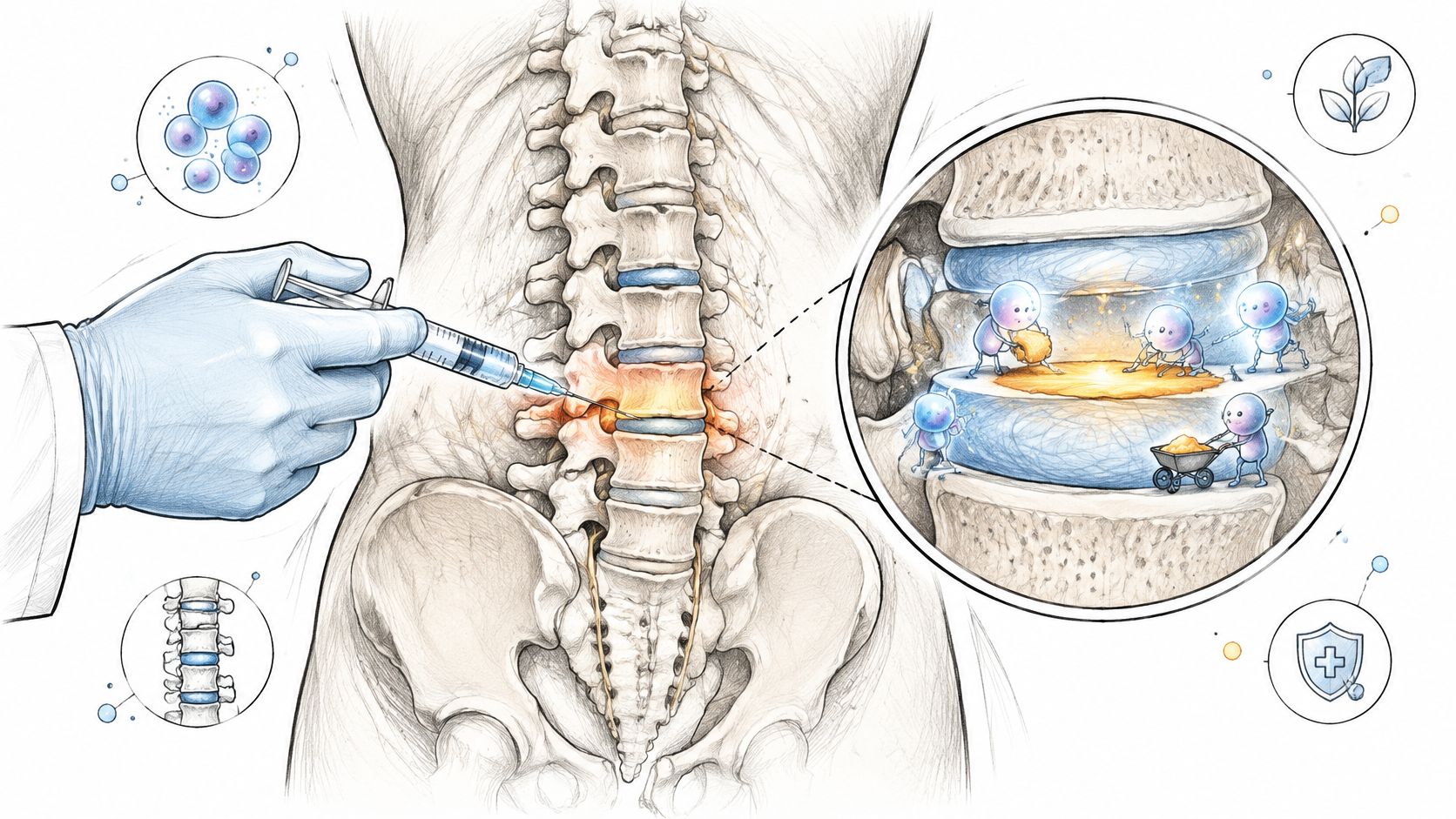
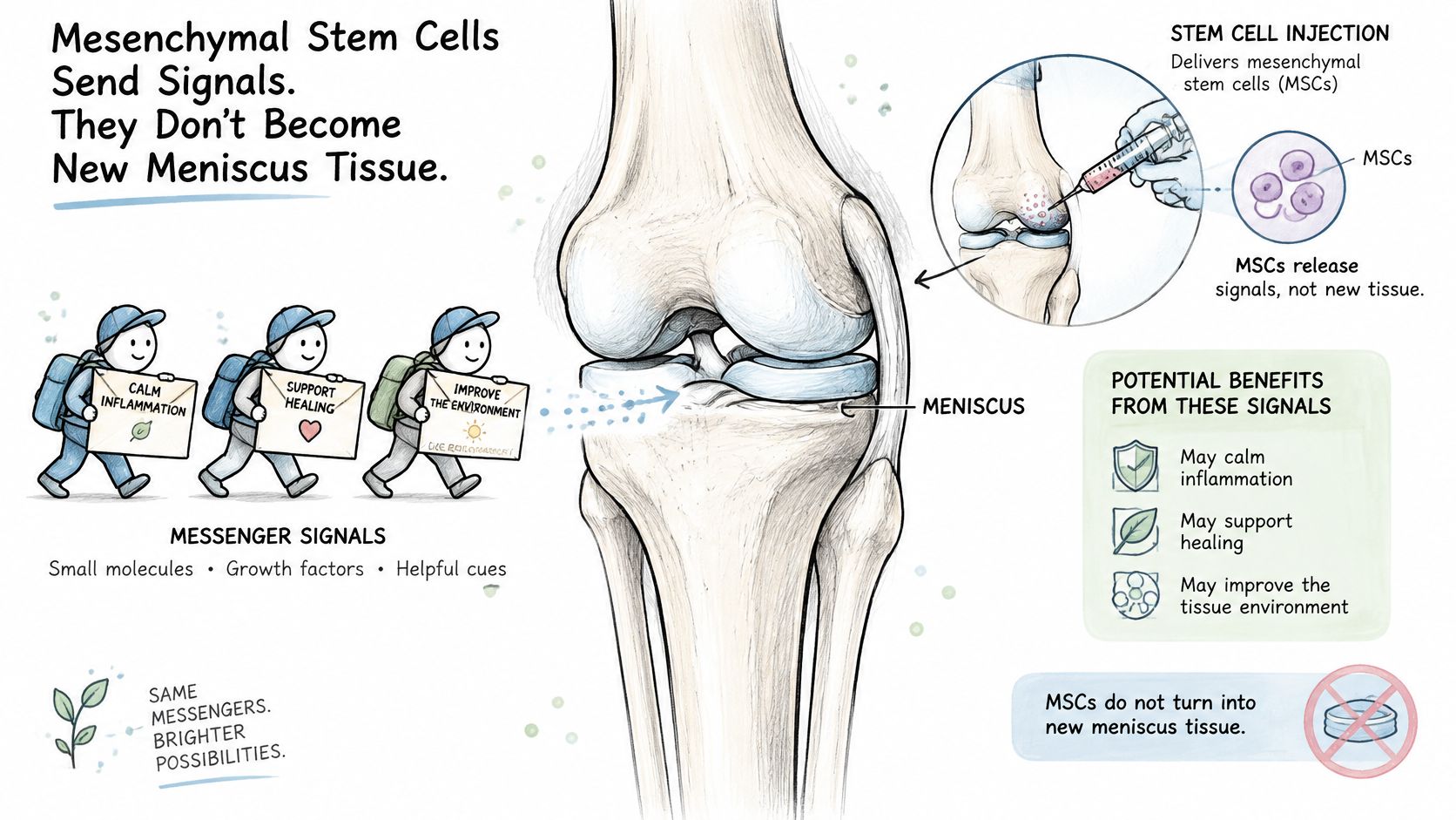
Think of mesenchymal stem cells as messengers rather than replacement building blocks. The proposed therapeutic action centers on the paracrine effect, the release of specialized signals that influence inflammation and local repair activity.
It's misleading to picture injected cells as becoming a new meniscus. MSCs have differentiation capacity under some conditions, but successful differentiation into replacement meniscal tissue isn't an established explanation for these clinical treatments. The practical discussion should focus on signaling, not a promise that injected cells transform into the missing structure.
What the positive clinical findings show
A first-in-human study of complex degenerative medial-meniscus tears reported improved symptoms and function in five men with a mean age of 48.2 years, following surgical repair plus synovial MSC transplantation, as detailed below.
A separate review of cell-based meniscal treatments summarizes the bone-marrow MSC and supported-repair results in the remaining rows; it also reports no increased meniscal volume in the control group of the volume study.
Reported stem cell results for meniscal injury
| Study group | Cells used | Reported result |
|---|---|---|
| Five men, mean age 48.2, surgical repair plus MSC transplant | 3.2 × 10^7 to 7.0 × 10^7 cells | Improved symptoms and function, followed to two years |
| Bone-marrow MSC meniscal study, 12-month MRI | 50 million cells | Increased meniscal volume in 24% of patients |
| Bone-marrow MSC meniscal study, 12-month MRI | 150 million cells | Increased meniscal volume in 6% of patients |
| MSC-supported repair series, five patients | Dose not specified in the summarized findings | Three of five had positive outcomes beyond 12 months, with MRI evidence of repair |
These are encouraging findings, but the studies used different cell sources and treatment methods. The complex-tear study combined cells with surgery, so it doesn't establish that injection alone produces the same result.
How to interpret an injection proposal
PRP supplies concentrated platelets and their signaling factors rather than replacement meniscus tissue. Neither PRP nor MSC signaling mechanically anchors a displaced fragment.
Our explanation of stem cell injections for knee pain provides context for a case-review discussion. That discussion should distinguish relief of inflammation, improved function, MRI changes, and restoration of a stable meniscus.
Josh's pre-procedure MRI showed a partially torn ACL and multiple meniscus tears. Our documented follow-up MRI nine months after treatment showed healing, but an individual outcome doesn't establish how another complex tear will respond.
Regulatory oversight also needs precise wording. Treatment within Mexico's COFEPRIS-regulated framework shouldn't be interpreted as proof of efficacy or as confirmation that a particular cell product is approved for meniscus repair; request documentation of the applicable authorization.
A promising imaging change isn't the same as proof that every tear has regained normal strength.
Recovery Timeline After Repair or Regenerative Care
Early protection after repair comes first. The surgeon's written limits govern knee bending, loading, and brace use, because rehabilitation must protect the specific repair rather than follow a generic exercise calendar.
Repair protocols commonly restrict flexion and weight-bearing during the initial six weeks. Physical therapy remains relevant after both surgical and nonoperative care, but starting therapy doesn't mean immediately loading the knee without restrictions.
Build capacity in stages
During protected rehabilitation, arrange practical support before you need it:
- Walking: Confirm whether you need crutches and how much weight you may place through the leg.
- Work and flights: Discuss prolonged sitting, airport distances, luggage, and individualized travel precautions.
- Driving: Obtain clearance based on braking ability, the affected leg, medication, and any brace.
- Exercise: Avoid deep bending, pivoting, and resistance beyond your prescribed limits.
During progressive rehabilitation, the therapist advances movement, strength, and walking according to swelling, control, and the procedure performed. A calendar date alone doesn't establish readiness for sport.
After an injection, pain relief and structural recovery may follow different timelines. The knee injection recovery guide can support planning, but improvement shouldn't automatically trigger unrestricted activity or routine repeat MRI without a clinical reason.
Once cleared, general advice on how to rebuild fitness with Empire Athletics can help with a gradual return to exercise. Your clinician's restrictions take precedence.
Establish the follow-up plan
A displaced tear, root injury, major instability, or rapidly worsening symptoms needs evaluation before an injection decision. Delaying a needed mechanical treatment can change the options available.
Bring these questions to your clinician:
- What movement and weight-bearing limits apply?
- When will the knee be imaged again, if repeat imaging is indicated?
- Which symptom means I should call immediately?
Choose Your Next Step for a Complex Tear
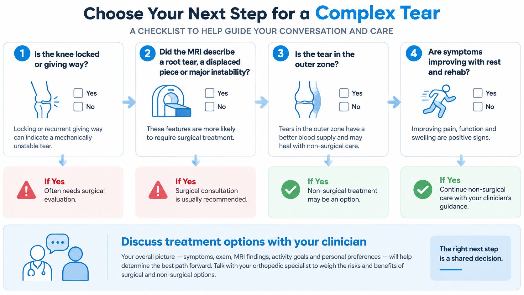
Use your MRI report to start a decision conversation, not to choose a treatment on your own. The most useful questions connect anatomy with what your knee can do.
- Is the knee locked or repeatedly giving way? A physical block or major instability moves the decision toward prompt orthopedic assessment.
- Does the report describe a root tear or displaced fragment? Ask specifically whether the attachment or moving tissue needs repair.
- Is the tear peripheral with viable tissue? That can make a preservation discussion particularly important.
- Are symptoms improving with rehabilitation? Track walking, swelling, and daily function rather than pain alone.
- Has someone recommended removal immediately? Ask why repair isn't suitable and what tissue would remain.
- What's your goal? Returning to pivoting sport, managing desk work, and improving comfortable walking can require different plans.
Where a regenerative consultation fits
Dream Body Clinic in Bucerias, Nayarit, Mexico, offers a stem cell knee treatment for selected knee conditions, including partial meniscal injuries. Our orthopedic treatments use MSCs with the aim of supporting repair in damaged cartilage, ligaments, and tendons, but a complex tear requires individual screening rather than an assumption of suitability.
Our knee protocol uses 50 million mesenchymal stem cells and includes an MRI. Joint regeneration treatment for a knee, hip, elbow, shoulder, or ankle costs $3,500 USD. A published cell count and price explain what's offered, not whether the treatment is appropriate for your tear.
We provide a complimentary case review and English-speaking coordination for international patients. Before making travel arrangements, send the complete MRI report, available images, relevant clinical notes, and a description of any locking or giving way.
Ask for a reasoned recommendation
A useful review should explain which findings support treatment and which make it unsuitable. It should also identify when an orthopedic surgical opinion is needed before considering regenerative care.
Decision standard: Choose a recommendation that explains what treatment can change, what it can't change, and how the clinician will assess your progress.
Ask how the team will distinguish improved comfort from structural healing, what rehabilitation you'll need, and what happens if symptoms persist. If the answer doesn't address displacement, root involvement, cartilage condition, and instability, the assessment isn't complete enough to guide a confident decision.
The right next step may be rehabilitation, a repair discussion, or a regenerative consultation. What matters is choosing it because of your knee's specific findings, not because complex sounds frightening or an injection sounds simpler.
Dream Body Clinic offers MRI-informed assessment and regenerative knee care for selected partial meniscal injuries. Visit Dream Body Clinic to request a complimentary case review, and send your full MRI report and clinical notes so our team can explain which features make your tear suitable or unsuitable for treatment.