
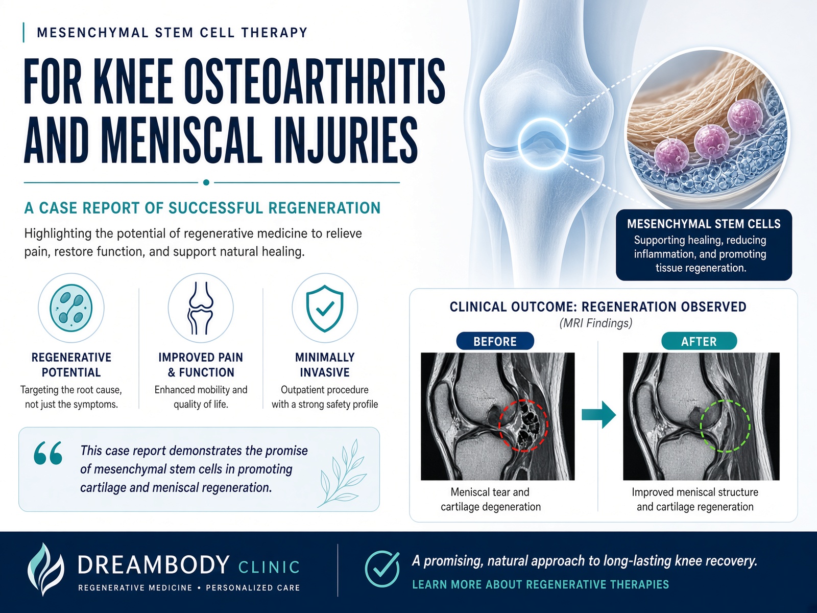
Mesenchymal Stem Cell Therapy for knee osteoarthritis and meniscal injuries- a case report of successful regeneration
September 01, 2026
Jaqueline Vega Trejo, MD1
Dream Body Clinic. Carmen Serdan 22, Bucerias, Nayarit 63732, Mexico.
Keywords: case report; mesenchymal stem cells; knee osteoarthritis; meniscus tear.
Case Summary:
A 45-year-old male presented with chronic left knee pain. Baseline MRI demonstrated horizontal tear involving the posterior horn of the medial meniscus, a grade I lesion of the lateral meniscus at the level of the posterior horn, grade I retropatellar osteoarthritis, grade II femorotibial osteoarthritis, and reactive synovitis. The patient received an ultrasound-guided intra-articular injection of 50 million mesenchymal stem cells derived from Wharton’s Jelly, followed by a structured rehabilitation program. At 6 months and 20 days post-treatment, follow-up MRI showed resolution of horizontal tear involving the posterior horn of the medial meniscus and the gonarthrosis (the new MRI report demonstrates preserved femorotibial and patellofemorotibial cartilage thickness without osteochondral defects) although the lesion of the lateral meniscus persists. Clinical symptoms and knee function also improved, suggesting a favorable response to treatment.
Introduction
Cartilage is the tissue that cushions the joints, allowing for smooth and pain-free movement.
Osteoarthritis is a chronic degenerative joint disease characterized by an inflammatory component, associated with degeneration and progressive loss of hyaline cartilage and subchondral bone, as well as damage to the synovial tissue, thickening and sclerosis of the subchondral plate, osteophyte formation, distention of the joint capsule, and changes in the periarticular soft tissues. MSCs can help to regenerate cartilage tissue by guiding chondrocytes, which are the cells that make up cartilage.
When MSCs are injected into the joint, they can reduce inflammation and promote the growth of new cartilage cells, leading to improved joint function and reduced pain.
The meniscus is a C-shaped piece of cartilage that acts as a shock absorber between the thigh bone and the shinbone.
Meniscal injuries of the knee include tears and ruptures of the meniscus, which generally occur following a rotational mechanism of the knee while the affected limb is weight-bearing and partially flexed. This may explain why the medial meniscus is involved 5 to 7 times more frequently than the lateral meniscus. Acute varus or valgus positioning of the knee can also cause meniscal tears. The diagnosis of meniscal injuries is primarily clinical and is based on the history of injury and sports participation, as well as the patient’s reported functional status and physical examination. MSCs can help to repair meniscus damage by promoting the growth of new meniscal tissue and reducing inflammation.
Clinical Case
A 45-year-old male patient presented with a 10-year history of intermittent left knee pain, localized to the anterior and medial areas, with a pain intensity of 4/10 on the Visual Analog Scale (VAS). He reports swelling and instability while walking, with symptoms worsening when climbing stairs and during prolonged periods of ambulation. Moderate limitation of flexion, extension, and internal and external rotation is noted. No crepitus is present.
The patient has undergone physical therapy with slight improvement. A corticosteroid injection was administered 10 years ago without improvement.
Medical History: The patient denied any chronic degenerative diseases, current medical treatment, no significant previous surgeries, or medication allergies.
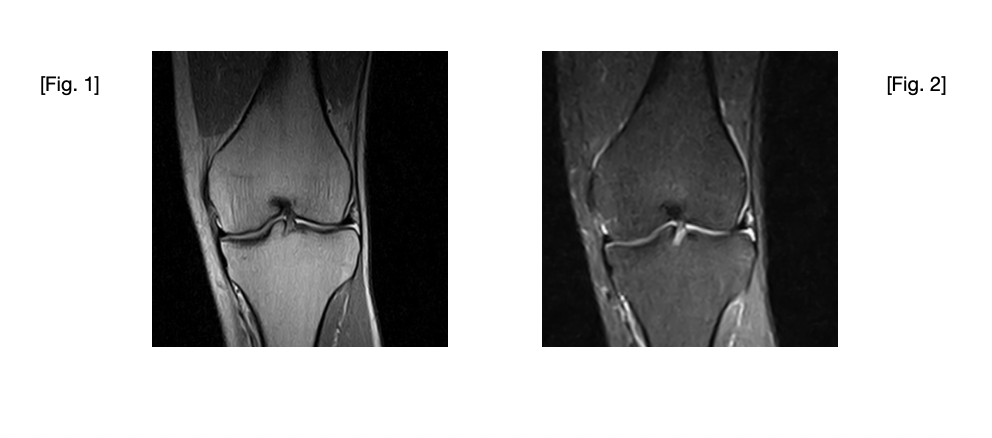
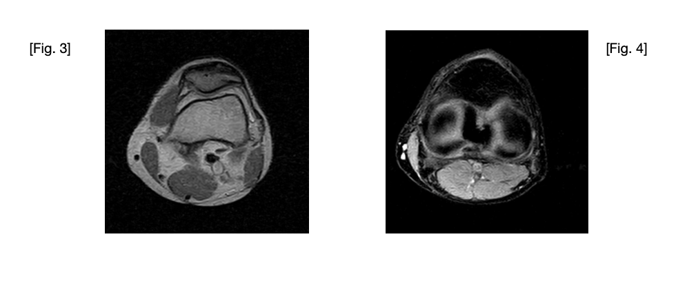
(2025-04-15) Left knee magnetic resonance imaging. Coronal Proton Density weighting (Fig. 1) and Coronal STIR (Fig. 2) sequence that shows: grade II femorotibial gonarthrosis and joint effusion suggesting reactive synovitis. (Fig. 3) Axial Proton Density weighting sequence: Wiberg type I patella with grade I retropatellar osteoarthritis. (Fig. 4) Axial XBONE T2 sequence shows: medial meniscus with an image suggesting a horizontal tear at the level of the posterior horn. Lateral meniscus with increased signal intensity toward the posterior horn, suggesting a grade I intrasubstantial lesion.
Therapeutic Intervention
50 million of human mesenchymal stem cells derived from Wharton Jelly (umbilical cord tissue) were applied via ultrasound guided intra-articular injection, with prior asepsis and antisepsis of the area. No adverse effects were reported by the patient during or after treatment.
After treatment the patient took 2 weeks off from exercise or sports. Hen then followed a 4 week physical rehab protocol provided during treatment. This is a protocol of physiotherapy exercises developed by our orthopedic surgeon to facilitate recovery. After the 6 weeks of rest and rehab the patient was able to return to his normal activities and exercise routine, we also recommended avoiding using any non
steroidal anti-inflammatory drugs (NSAIDS), Prednisone, or anti-inflammatory supplements such as gingko biloba or turmeric for at least 2 weeks post treatment and preferably for 6 weeks post treatment, instead, the patient was prescribed with paracetamol/tramadol 37.5mg/325mg 1 pill every 8 hours just in case of pain.
Follow-up
During his second visit, the patient reported improvement in pain; intermittent, appearing with movement, slight limitation of movement and range of motion.
A second MRI was taken 6 months and 20 days after treatment with the following results:
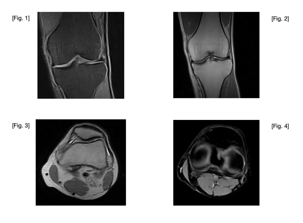
(2025-11-04) Left knee magnetic resonance imaging. Coronal SPED ( Fig. 1) and Coronal Proton Density (Fig. 2) weighting shows: articular cartilage of the medial and lateral femorotibial compartments preserved in thickness, with no osteochondral defects. (Fig. 3) Axial Proton Density weighting sequence: Patella and Patellofemoral joint: Wiberg type I patella, with preserved cartilage thickness. (Fig. 4) Axial XBONE T2 sequence shows: lateral meniscus increased signal intensity in the posterior horn, suggestive of a grade I intrasubstance lesion.
Results
Baseline MRI of the left knee demonstrated a horizontal tear involving the posterior horn of the medial meniscus, a grade 1 lesion of the lateral meniscus at the level of the posterior horn, grade 1 retropatellar osteoarthritis, grade II femorotibial osteoarthritis, and reactive synovitis.
Follow-up MRI, performed after mesenchymal stem cell infiltration, revealed persistent Grade 1 injury of the lateral meniscus. No findings suggestive of structural deterioration were identified.
Comparison of the two MRI examinations demonstrated improvement in structural findings, most notably the resolution of the previously reported horizontal tear involving the posterior horn of the medial meniscus and the previously reported osteoarthritis. The new MRI report demonstrates preserved femorotibial and patellofemoral cartilage thickness without osteochondral defects, although the lesion of the lateral meniscus persists.
Discussion
Knee osteoarthritis is a highly prevalent disease, associated with significant social and economic costs. Current evidence indicates that this condition is multifactorial, moving away from the concept of an exclusively degenerative disease and recognizing the importance of the inflammatory component. A torn meniscus is one of the most common knee injuries. A torn meniscus may result in an inability to move the knee or persistent pain.
MSC treatment is an alternative that has shown promising results for meniscal injuries and cartilage regeneration.
The administration of 50 million mesenchymal stem cells via intra-articular injection is minimally invasive and associated with a faster recovery period and minimal side effects. Compared with surgical procedures, MSC therapy may allow patients to return to daily activities more quickly while demonstrating encouraging results in meniscal injuries and cartilage regeneration.
Strengths/limitations: conclusions are limited by the single-patient design and the absence of a control group.
References
- Mautner K, Gottschalk M, Boden SD, et al. Cell-based versus corticosteroid injections for knee pain in osteoarthritis: a randomized phase 3 trial. Nat Med. 2023;29(12):3120-3126. doi:10.1038/s41591-023-02632-w.
- Instituto Mexicano del Seguro Social. Diagnóstico y Tratamiento de las Lesiones de Meniscos en Rodilla del Adulto. Guía de Práctica Clínica GPC; IMSS-577-12. Instituto Mexicano del Seguro Social; 2012.
- Instituto Mexicano del Seguro Social. Prevención, Diagnóstico y Tratamiento de Rehabilitación en el Paciente Adulto con Osteoartrosis de Rodilla en los Tres Niveles de Atención. Guía de Práctica Clínica GPC; IMSS-726-14. Instituto Mexicano del Seguro Social; 2014.
- American Academy of Orthopaedic Surgeons. Management of Osteoarthritis of the Knee (Non-Arthroplasty): Evidence-Based Clinical Practice Guideline. 3rd ed. American Academy of Orthopaedic Surgeons; 2021.
- American Academy of Orthopaedic Surgeons. The Management of Osteoarthritis of the Knee (Surgical and Non-Surgical). American Academy of Orthopaedic Surgeons.
- Whittle SL, Johnston RV, McDonald S, et al. Stem cell injections for osteoarthritis of the knee. Cochrane Database Syst Revt 2025.
Learn All About the Stem Cell Knee Treatment by Clicking Here
Call (888) 704-3977 or WhatsApp +523222321055 for a Free Consultation