

The most popular advice about herniated disk stem cell treatment is also the most misleading: injected cells will become new disc tissue, rebuild the annulus, and make the herniation disappear. That isn't what current human evidence establishes. Mesenchymal stromal cells, often called MSCs, appear to work mainly through a paracrine effect, releasing specialized signals that influence the patient's own disc cells rather than reliably differentiating into replacement disc cells.
That distinction matters because a painful disc can involve two different problems. A herniated fragment may mechanically irritate or compress a nerve, while degeneration may create chronic discogenic pain through inflammation and loss of healthy matrix. MSC therapy is being investigated mainly for the second biology, not as a dependable way to remove a large fragment or urgently decompress a nerve.
Table of Contents
- Why a Herniated Disc Needs More Than Mechanical Relief
- The Paracrine Effect and How Stem Cells Actually Signal Repair
- Delivery Routes for Reaching a Damaged Disc
- Who Is a Good Candidate for Stem Cell Treatment
- A Realistic Patient Journey From MRI to Follow-Up
- What the Evidence Actually Shows in 2026
- Risks, Contraindications, and Questions to Ask Any Clinic
- What Realistic Results Look Like and What Comes Next
Why a Herniated Disc Needs More Than Mechanical Relief
A disc has a tough outer ring, the annulus fibrosus, and a softer inner region, the nucleus pulposus. When the inner material pushes through a weakened outer ring, the result can be a herniation. If that material contacts a nerve root, the patient may develop sciatica, numbness, tingling, or weakness. Degenerative disc disease is related, but it isn't identical. It describes deterioration of the disc environment and can cause back pain even without an acute nerve-compressing fragment.
Conventional treatment often focuses on reducing symptoms or relieving pressure. Physical therapy, medication, activity modification, and targeted injections may help calm inflammation and restore movement. Surgery can be appropriate when a structural lesion causes significant or progressive neurological compromise. None of these approaches automatically answers a different question: can the damaged disc environment become less inflammatory and more supportive of its resident cells?
Why the disc is difficult to repair
Disc tissue has limited blood supply, especially in the regions commonly involved in degeneration and annular injury. That restricted circulation helps explain why discs don't repair themselves as readily as tissues with a richer blood supply. It also creates a difficult setting for any injected therapy. A cell product must survive, interact with local tissue, and influence the inflammatory and matrix signals without causing additional injury.
The regenerative-medicine hypothesis begins here. MSCs may release molecules that alter the behavior of nucleus-pulposus and annulus-fibrosus cells. They are not microscopic replacement parts. They act more like temporary biological coordinators.
Practical rule: Pain relief, nerve decompression, and structural disc repair are separate outcomes. A treatment can influence one without proving the others.
The phrase paracrine signaling describes this process. An MSC releases signals into its surroundings, and those signals may reduce damaging inflammation, influence matrix turnover, and encourage local cells to survive or function differently. The cells don't need to transform into new disc cells for that communication to occur.
That is why a careful discussion of treatment must separate discogenic pain from mechanical nerve compression. A contained degenerative disc with chronic back pain may be considered for investigational MSC treatment after conservative care. A large extruded fragment causing progressive weakness or cauda-equina symptoms needs prompt spine-specialist assessment, not a promise that cells will dissolve the obstruction.
The Paracrine Effect and How Stem Cells Actually Signal Repair
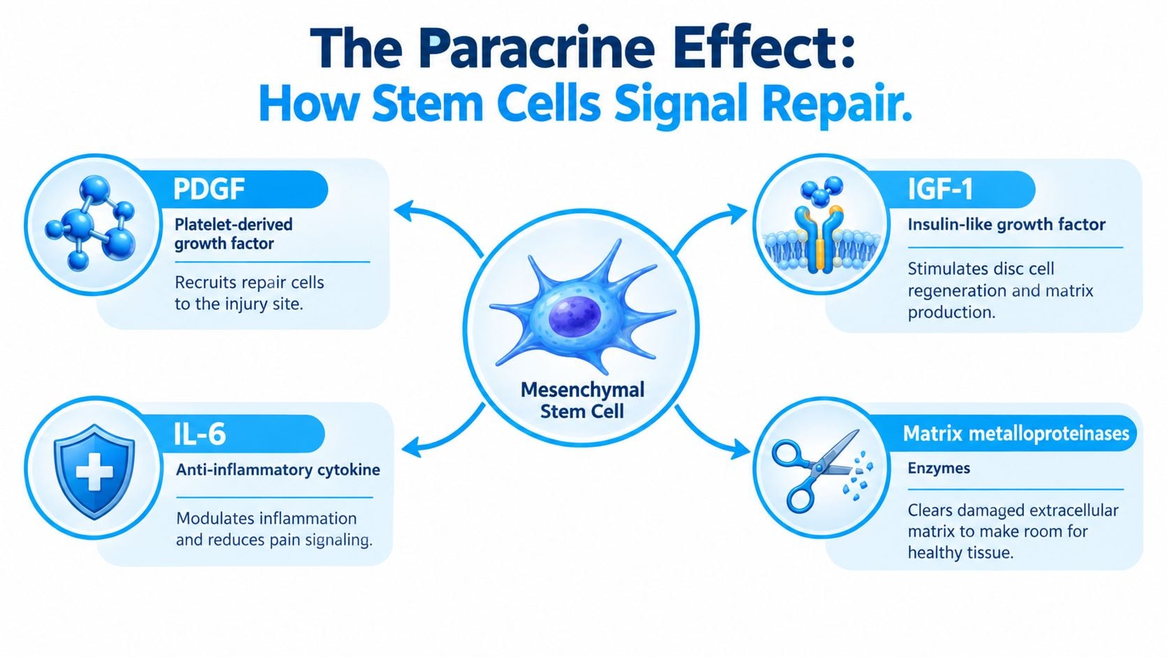
Think of an MSC injection as a biological messaging system, not a shipment of replacement cartilage. The cells can release soluble factors that interact with resident nucleus-pulposus and annulus-fibrosus cells. A review of MSC-derived exosomes describes this signaling model and identifies factors including platelet-derived growth factor, or PDGF, insulin-like growth factor-1, or IGF-1, interleukin-6, or IL-6, and matrix metalloproteinases (review of MSC-derived exosomes and disc repair signaling).
What the signals may change
PDGF can participate in communication that recruits and activates repair-related cells. IGF-1 may influence cell survival and extracellular-matrix production. IL-6 is more complicated than a simple “anti-inflammatory” label, because its effects depend on context, concentration, and signaling pathway. In disc research, MSC-mediated immune and inflammatory modulation is part of the proposed therapeutic effect, but patients shouldn't be told that one cytokine guarantees pain relief.
Matrix metalloproteinases are enzymes involved in extracellular-matrix turnover. That doesn't mean they "cut out" a herniation. It means they may participate in remodeling damaged matrix, alongside other signals that influence resident disc cells. The hoped-for result is a more favorable local environment, not a guaranteed anatomical reconstruction.
MSC-derived exosomes add another layer. These extracellular vesicles carry proteins, nucleic acids, and other molecular messages that can change the behavior of nearby cells. Experimental work has associated these signals with increased nucleus-pulposus-cell proliferation, lower cell death, altered inflammatory pathways, and improved matrix-related activity. A review of exosome research describes animal findings involving slower degeneration, reduced cellular senescence, and preservation of disc height, but those findings remain largely preclinical (review of MSC exosome mechanisms in intervertebral-disc treatment).
One especially useful experiment tested whether exosome release mattered. Researchers used GW4869, an inhibitor of exosome secretion. When exosome release from MSCs was blocked, the protective effect against pyroptosis in nucleus-pulposus cells was abolished. That result supports the central point of this guide: the benefit depends substantially on what MSCs release, not on the cells reliably becoming replacement disc tissue.
Research also links MSC signaling with fibrosis and resident-cell activity. Studies describe paracrine effects on nucleus-pulposus cells, regulation of profibrotic pathways, and stimulation of local progenitor populations such as Tie2-positive cells (research on MSC homing and paracrine stimulation). These mechanisms are biologically plausible, but laboratory and animal findings don't prove that a person's herniated disc will heal.
For a patient-friendly explanation of the signaling concept, this overview of how stem cell injections work can be a useful starting point. The key question remains clinical: does the communication produce durable improvement in the right patient, with acceptable risk and objective follow-up?
Delivery Routes for Reaching a Damaged Disc
The injection route determines which tissue receives the greatest exposure. It also determines what the treatment can realistically target. An epidural injection may reach inflamed structures around a nerve root, while an intradiscal injection places the product inside the disc environment. Intravenous and intrathecal approaches have broader or different targets and shouldn't be described as interchangeable with direct disc treatment.
A practical comparison
| Route | Target | Common Dose | Best For |
|---|---|---|---|
| Intradiscal | The disc space, usually the nucleus-pulposus region or selected disc level | Protocol-dependent | Investigational treatment for discogenic pain and degeneration |
| Epidural | The epidural space and nearby inflamed nerve root | Protocol-dependent | Radicular inflammation when the main target is around the nerve |
| Intravenous | The circulation and systemic immune environment | Protocol-dependent | Broader immune modulation, not precise disc delivery |
| Intrathecal | Cerebrospinal fluid around the spinal cord and nerves | Protocol-dependent | Selected neurological indications, not routine direct disc repair |
Intradiscal delivery is the most direct route for studying disc biology. It can place MSCs close to resident disc cells, but it doesn't mechanically remove a fragment or guarantee that the cells remain active. Epidural delivery may be more logical when inflammation around a nerve root dominates the symptoms, although it doesn't equal direct treatment of disc degeneration.
Intravenous infusion exposes the body to a wider cellular and immune environment. It may be considered when a protocol aims at systemic immune modulation, but it can't be assumed to deliver a therapeutic concentration to one specific lumbar disc. Intrathecal injection places cells or biological material into cerebrospinal fluid and carries a different risk profile. It is generally associated with selected neurological protocols rather than routine treatment of an isolated herniated disc.
Patients comparing routes can review osteopathic injection therapy options to understand how injection-based care may be framed across musculoskeletal conditions. For disc-specific context, this discussion of stem cell treatment for degenerative disc disease may help clarify why route and diagnosis have to match.
A clinic should explain the exact target, imaging method, product, dose, and reason for choosing a route. “Stem cells for back pain” is too broad to support informed consent.
Who Is a Good Candidate for Stem Cell Treatment
The most plausible candidate is not anyone with an abnormal MRI. A useful evaluation starts with the pain pattern, neurological examination, imaging, and response to conservative care. MSC treatment remains investigational, so the patient should understand both the potential rationale and the limits of the evidence.
Features that may support consideration
Patients may be evaluated when they have:
- Chronic discogenic low-back pain: Symptoms appear to arise from a degenerative disc rather than only from muscle, facet joints, or the sacroiliac joint.
- MRI-confirmed disc pathology: Imaging identifies the affected level, degeneration pattern, disc height, and any nerve-root contact.
- Persistent symptoms after conservative care: Physical therapy, activity changes, medication, or other nonoperative measures haven't provided adequate relief.
- Contained herniation or degeneration without urgent neurological decline: The anatomy doesn't demand immediate decompression.
MRI matters because a bright or dark disc on a scan doesn't explain the entire pain experience. A specialist should compare the imaging level with the clinical examination and consider competing pain generators. The Pfirrmann grade, annular changes, disc height, nerve-root contact, and symptoms all influence the decision.
When regenerative care should not delay specialist treatment
Progressive motor weakness, new bowel or bladder changes, saddle anesthesia, or a large extruded fragment require urgent assessment by a spine specialist. Severe fixed nerve compression is a mechanical problem that an injection may not relieve. A patient shouldn't let the availability of an appealing biological treatment postpone evaluation for a potentially time-sensitive condition.
Pregnancy, active infection, known malignancy concerns, major bleeding risk, and severe instability also require careful medical review. The right question isn't “Can MSCs help any back pain?” It's “Does the proposed mechanism match the dominant pain generator, and is it safe to investigate in this patient?”
A Realistic Patient Journey From MRI to Follow-Up
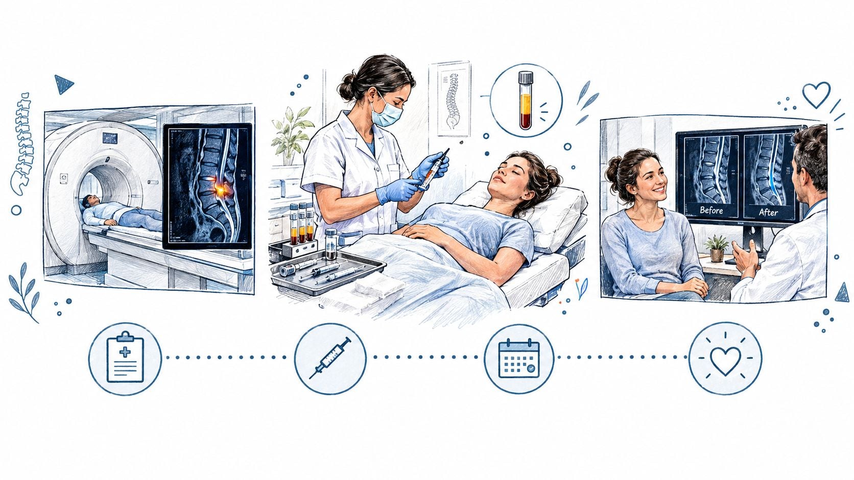
A responsible regenerative-spine pathway begins before anyone discusses an injection. The clinic reviews symptoms, neurological findings, prior treatment, medications, and MRI images. The purpose is to identify the level being treated and distinguish discogenic pain from nerve compression, facet pain, sacroiliac pain, or another cause.
From planning to procedure
A clinician may recommend baseline MRI documentation and define what will be measured later. Useful outcomes include pain intensity, walking tolerance, sitting tolerance, sleep, medication use, neurological symptoms, and a validated disability score. A post-treatment MRI can show whether imaging changed, but an unchanged scan doesn't automatically mean a patient has failed clinically, and a changed scan doesn't prove nerve decompression.
Some protocols pair MSC treatment with platelet-rich plasma, or PRP, as an adjunct intended to add signaling molecules to the local environment. That combination is protocol-specific and shouldn't be treated as a universal requirement. The procedure itself may involve image-guided access to one or more lumbar levels, monitoring, and a period of observation before discharge.
Published protocols vary in cell source, preparation, injection technique, and dose. The clinic information supplied for orthopedic and spine protocols describes ranges such as 25 to 100+ million MSCs, but dose alone doesn't predict an outcome. Cell identity, viability, potency, sterility, delivery accuracy, patient selection, and the condition of the disc all matter.
A typical care plan may include structured remote check-ins at approximately three, six, and twelve months, with instructions about activity, rehabilitation, warning symptoms, and when to repeat imaging. Early soreness can occur after an injection, while meaningful changes may take time. The patient should know who handles urgent concerns after returning home.
Ask before treatment: What will be measured, when will it be measured, and what finding would change the plan?
The commitment is therefore larger than the injection appointment. It includes diagnostic review, informed consent, recovery guidance, rehabilitation, symptom tracking, and follow-up that can distinguish temporary pain reduction from durable functional improvement.
What the Evidence Actually Shows in 2026
The human evidence is promising enough to justify continued research, but not strong enough to support claims of a reliable cure. A 2023 systematic review and meta-analysis identified 2,392 records and included only 9 eligible studies involving 245 patients. Among them, 193 patients received mesenchymal stem-cell injections. Pain measured with a visual analogue scale improved significantly, with a reported mean difference of 41.62 points and a 95% confidence interval of 24.32 to 58.93 (2023 systematic review and meta-analysis).
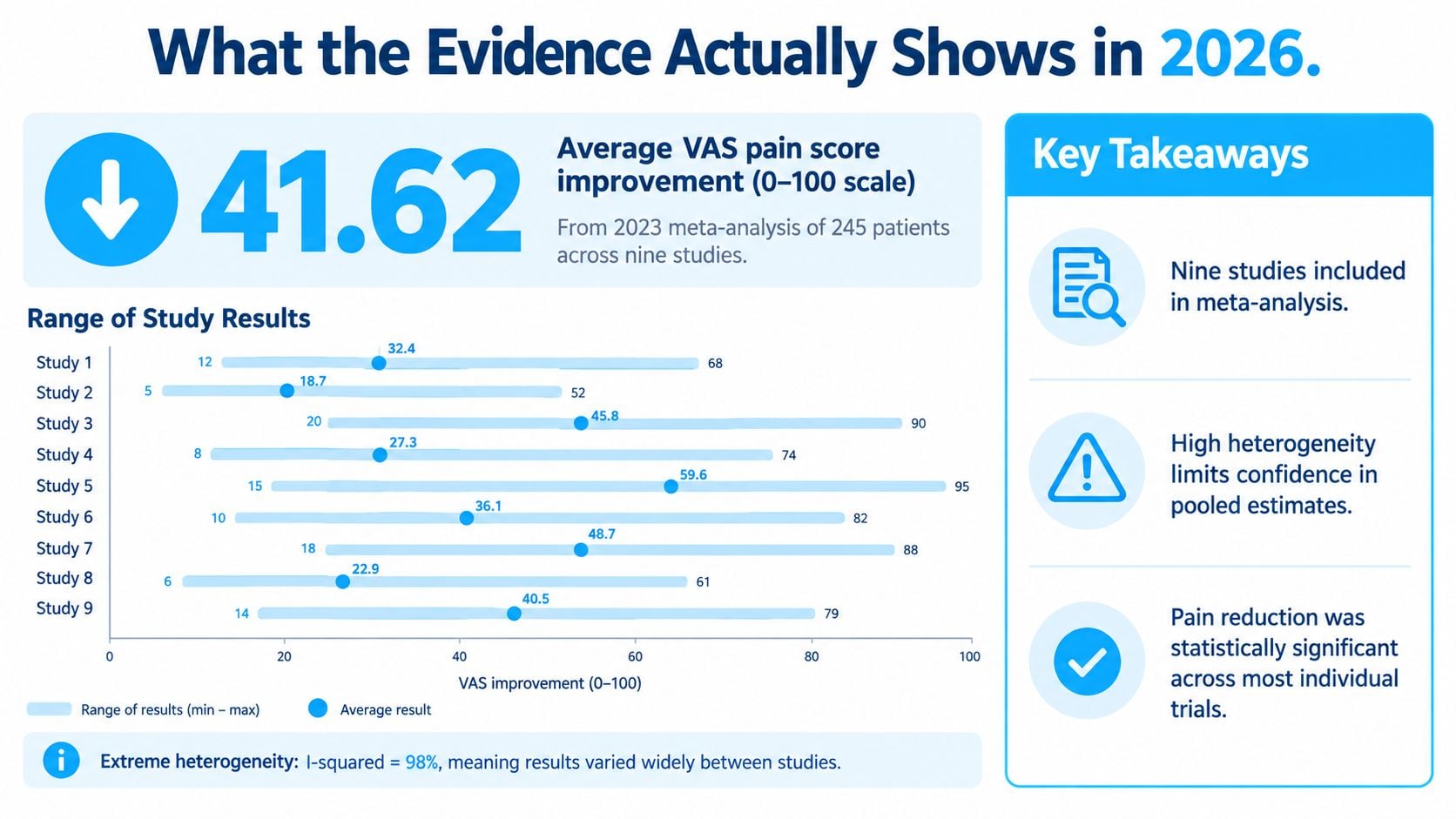
That result needs context. Statistical heterogeneity was extremely high, with I² = 98%, meaning the studies differed substantially in patient selection, cell products, injection methods, comparators, and outcome measurement. The same review reported improvements in the Oswestry Disability Index and suggested potentially lower adverse-event and reoperation rates, but it didn't establish that treatment reverses a herniation or reliably regenerates disc tissue.
Why newer reviews remain cautious
A 2025 systematic review reported 13 clinical studies published from 2011 through 2025, totaling 1,299 participants. Only 5 studies were randomized controlled trials, while 8 were prospective, retrospective, cohort, or pilot studies. The review found intradiscal treatment appeared generally safe in the short to medium term and produced modest improvements in pain and disability, but it found no compelling imaging evidence of clear biological disc repair or a proven disease-modifying effect (2025 systematic review of clinical MSC studies).
Earlier evidence shows why surgery rates shouldn't be presented casually. A 2019 review found that the proportion of patients requiring subsequent surgery ranged from 6% to 80%, reflecting major differences in study populations and protocols. A separate 2023 review located 11 clinical studies but only 1 randomized controlled trial, concluding that available human evidence didn't support routine use.
A 2025 randomized phase IIb report found autologous bone-marrow MSC injection to be safe and possibly associated with slight structural improvement, yet it didn't show a clear six-month advantage over a sham procedure for pain, disability, or quality of life. A separate phase-I study treated only five patients with 9 million allogeneic bone-marrow clonal MSCs, and no serious adverse events occurred, but the authors emphasized the need for larger cohorts and longer follow-up (2025 clinical evidence review and trial discussion).
For clinicians comparing supportive approaches, this guide to HBOT for healthcare professionals offers context on another therapy discussed in back-pain care. It shouldn't be confused with evidence that MSCs regenerate a disc.
The evidence supports investigational potential and symptom improvement in selected patients. It doesn't prove anatomical healing, resolved nerve compression, or reliable avoidance of surgery.
Risks, Contraindications, and Questions to Ask Any Clinic
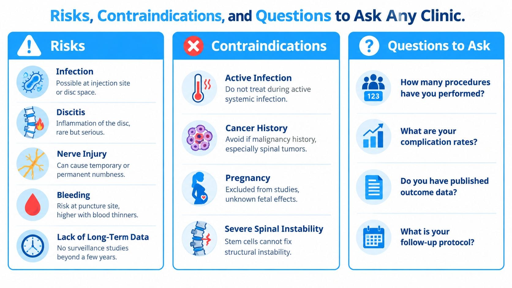
Short-term procedural safety is not the same as proven long-term regeneration. Risks can include infection, discitis, bleeding, nerve injury, worsening pain, and complications related to the injection route. Long-term surveillance remains limited, so a clinic shouldn't imply that an encouraging early safety profile proves decades of safety.
Medical screening comes first
Active systemic or local infection is a reason to postpone treatment. Active malignancy, particularly concern for a spinal tumor, requires specialist evaluation. Severe coagulopathy or unsafe anticoagulation can increase procedural risk, and pregnancy is generally avoided because fetal effects are unknown. Severe spinal instability is also outside what cell therapy can mechanically correct.
Patients should ask direct questions before agreeing to treatment:
- Cell identity: What cells are being used, and how are identity and potency assessed?
- Safety testing: What sterility, contamination, and endotoxin tests are performed?
- Procedure guidance: Will imaging guide the needle to the selected disc or target?
- Clinical fit: What finding makes this route and level appropriate for my symptoms?
- Retreatment policy: What would justify another injection, and what would make retreatment inappropriate?
- Follow-up plan: Who reviews outcomes, imaging, and complications after the patient returns home?
A clinic operating in Mexico may describe its treatment framework as being delivered under COFEPRIS oversight. That regulatory context is relevant for international patients, but it doesn't replace a discussion of the evidence for the specific cell product, indication, and procedure.
Patients should also plan for unexpected symptoms. New weakness, fever, severe escalating pain, drainage, or changes in bladder or bowel function need prompt medical attention. Guidance on worse pain after stem cell injection may help patients prepare questions, but it isn't a substitute for contacting a clinician when warning signs appear.
What Realistic Results Look Like and What Comes Next
Paracrine signaling explains why pain and function might improve even when an MRI looks similar. A calmer inflammatory environment can change how resident disc cells and nearby nerves behave without proving that the disc has been rebuilt. Likewise, a better pain score doesn't demonstrate that a herniation has anatomically healed.
Some patients may notice early changes over four to eight weeks, while further gains may develop over three to six months, but individual timelines vary and these periods aren't guarantees. Track function, walking, sitting, sleep, medication use, neurological symptoms, and objective follow-up rather than relying on pain alone.
A sensible checklist is short:
- Confirm the diagnosis and urgent red flags.
- Ask whether the target is discogenic pain or mechanical compression.
- Request details about cells, testing, dose, route, and imaging guidance.
- Compare sham-controlled evidence with uncontrolled reports.
- Secure a follow-up plan that extends beyond the early recovery period.
For conservative recovery planning and rehabilitation context, patients may also review disc recovery without surgery. Larger randomized trials, standardized cell preparations, objective MRI criteria, and longer follow-up are still needed before MSC therapy can be considered a proven regenerative cure.
Dream Body Clinic offers case review and regenerative spine protocols that may include image-guided MSC treatment, PRP, MRI planning, and structured remote follow-up for selected patients. If you're considering herniated disk stem cell treatment, visit Dream Body Clinic to submit your medical information and discuss whether the proposed approach matches your diagnosis.