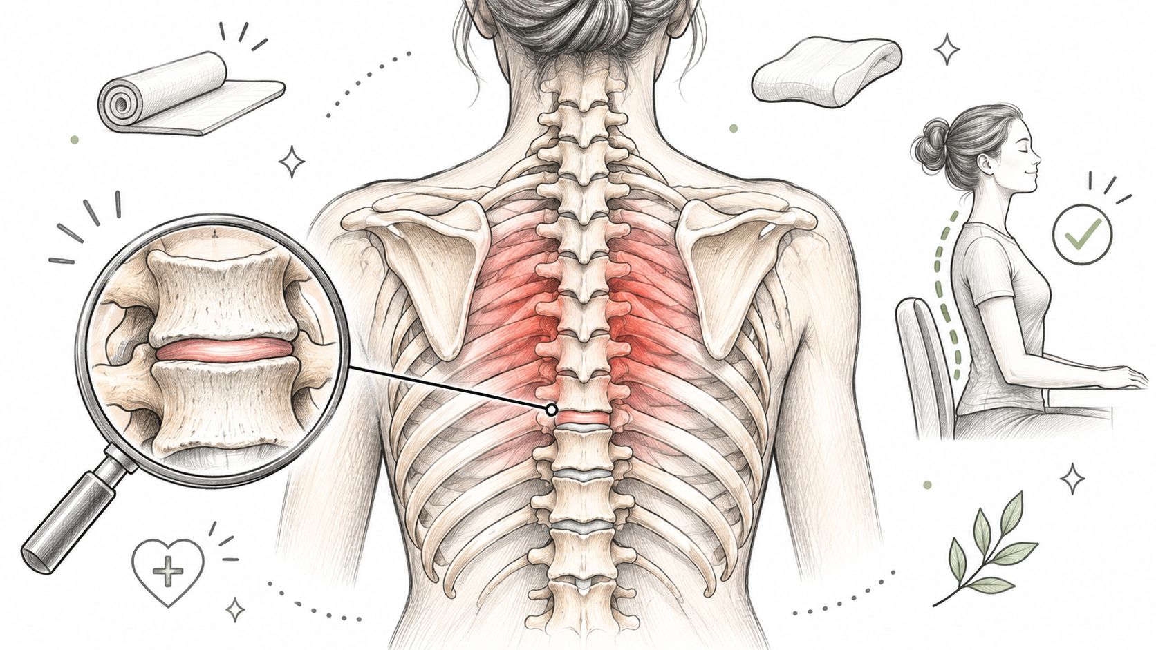
You've been sitting at your desk or driving when a deep ache starts between your shoulder blades. Your MRI report mentions degeneration, and now you're wondering whether the discs in your mid-back are wearing out, whether that explains the pain, and what treatment makes sense.
Degenerative disc disease of the thoracic spine isn't a diagnosis that an MRI can establish by itself. The useful starting point is to match your pain pattern and neurological examination to the scan, while checking for conditions that need a different kind of care.
Table of Contents
- Symptoms That Point to Thoracic Disc Degeneration
- Urgent Warning Signs to Rule Out First
- What Drives Thoracic Disc Wear Over Time
- How Doctors Confirm a Thoracic Disc Diagnosis
- Conventional Care Options and Their Limits
- Regenerative Stem Cell Options and What Studies Show
- Confirm Your Next Step Before Choosing Treatment
Symptoms That Point to Thoracic Disc Degeneration
Say your mid-back aches after reading with your head bent forward. Supporting your back helps, and lying down settles the discomfort. That pattern suggests a problem influenced by position and mechanical load, although it doesn't identify a disc as the cause.
The thoracic spine behaves differently from the neck and lower back because the rib cage helps stabilize it and limits movement. Disc-related symptoms are consequently less common here than in more mobile spinal regions. When symptoms do occur, they can involve the mid-back, ribs, or chest wall.
Typical thoracic disc symptoms and what they suggest
| Symptom pattern | Typical location | What it suggests |
|---|---|---|
| Dull, aching pain worse with sitting or bending | Between shoulder blades and lower ribs | Mechanical disc or facet-joint irritation |
| Band-like pain wrapping around the ribs | Chest wall or flank | Possible nerve root irritation; other causes must be excluded |
| Stiffness after prolonged sitting or on rising | Upper and mid back | Reduced segmental movement |
| Pain that changes with position and eases lying down | Mid-back | Load-related pattern compatible with degeneration |
| Pain with deep breathing, coughing or chest pressure | Chest wall or front of chest | Not typical of disc pain alone; needs cardiopulmonary evaluation |
Look for patterns rather than a single symptom
A dull central ache is different from burning pain that follows a narrow strip around the ribs. The first may involve discs, joints, or muscles; the second can suggest irritation of a spinal nerve. Tenderness when you press on the chest wall may involve local muscles or rib joints, but tenderness alone doesn't establish a harmless cause.
Stiffness after sitting or on rising can accompany reduced movement and muscle guarding. Notice whether gentle movement helps, whether a particular position reliably reproduces the pain, and whether symptoms interrupt sleep or persist at rest.
Practical self-check: Record where the pain sits, what makes it better or worse, how long stiffness lasts, and whether you notice numbness, weakness, or bladder or bowel changes.
Bring that log to your appointment rather than relying on memory. Don't deliberately provoke symptoms with heavy lifting or repeated twisting to test yourself. Pain with exertion, chest pressure, or breathing symptoms needs medical assessment rather than a home posture experiment.
Urgent Warning Signs to Rule Out First
Not all mid-back pain is disc wear. Thoracic disc disorders can produce chest-wall or upper-abdominal symptoms, but heart, lung, abdominal, and other conditions can also cause pain in this region, as the StatPearls overview of thoracic discogenic syndrome explains.
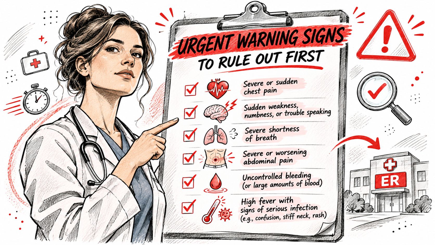
The safest response is to assess the warning signs before choosing an injection, exercise program, or treatment abroad. This checklist highlights symptoms that change the plan.
Red flags and the response each requires
| Warning sign | Why it matters | Response |
|---|---|---|
| Chest pressure, breathlessness or pain on exertion | Can signal cardiac or lung disease | Emergency care now |
| Fever, infection risk or unexplained weight loss | Can signal infection or malignancy | Urgent medical review |
| Cancer history or significant recent trauma | Raises risk of fracture or spinal lesion | Prompt imaging and specialist review |
| Osteoporosis or recent fracture | Raises risk of vertebral collapse | Medical review within the same week, sooner for new severe pain |
| Progressive leg weakness, gait change or bladder or bowel change | Can signal spinal-cord compression | Urgent spinal specialist review |
| Sensory change below a trunk level | Can signal cord or nerve involvement | Urgent spinal specialist review |
Distinguish back pain from spinal-cord symptoms
Axial pain means pain centered around the spine. Myelopathy means impaired spinal-cord function, which may affect walking, leg control, sensation, or bladder function rather than making the back hurt.
A new tendency to stumble, increasing leg stiffness, or a clear boundary below which sensation feels different deserves particular attention. Sudden or rapidly worsening weakness, inability to walk, or new loss of bladder or bowel control warrants emergency evaluation. An existing MRI doesn't replace a current neurological examination.
Our thoracic spine pain information can help you prepare questions, but remote review isn't an appropriate substitute for urgent local assessment.
Before seeking care abroad, gather:
- Imaging records: Prior MRI images and X-ray reports identifying the affected levels.
- Medication details: Current prescriptions, supplements, allergies, and blood thinners.
- Medical history: Cancer, infections, fractures, osteoporosis, and previous spinal procedures.
- Recent testing: Available blood tests and relevant specialist assessments.
Use emergency services for cardiopulmonary symptoms and urgent spinal assessment for cord signs. Don't postpone that care until a travel date or elective consultation.
What Drives Thoracic Disc Wear Over Time
Think of a disc as a water-rich cushion between vertebrae, a little like a jam-filled doughnut with a tough outer ring. The comparison isn't exact, but it helps explain how the softer center and fibrous outer layers share load. As discs lose hydration, they can become less resilient and lose height.
Age-related changes aren't the only consideration. Repetitive loading, prolonged forward-bent positions, earlier injuries, smoking that impairs tissue nutrition, and inactivity that reduces muscular support can contribute to the overall problem. These factors don't let a clinician calculate which disc hurts, but they help guide practical changes.
A structured assessment separates those contributing factors from the actual diagnosis.
What prevalence figures actually mean
A systematic review of thoracic degenerative MRI findings reported prevalence estimates ranging from 0.2% to 89%, reflecting different imaging definitions, ages, and recruitment settings. The same review summarized a study of 2,007 subjects in which 58.5% had thoracic degeneration, 2.3% of participants had isolated thoracic degeneration, and 96% of those with thoracic changes also had degeneration elsewhere in the spine.
In an older cohort summarized by that review, 420 adults, with mean ages of approximately 82 years, had disc protrusion reported in 31.5% of men and 37.4% of women. These are imaging findings, not estimates of how many people needed treatment.
The distinction matters when someone sees several abnormalities on a report. More widespread degeneration doesn't automatically mean more severe symptoms, and the most abnormal-looking disc isn't necessarily the painful one.
A scan describes structure. Your clinician still has to determine which findings, if any, explain your symptoms.
For a visual introduction to the clinic's spine-treatment discussion, you can watch the following video.

The practical response to disc wear is to improve tolerable movement, reduce unnecessary strain, and investigate the symptoms that limit your life. It isn't to assume every age-associated change needs an intervention.
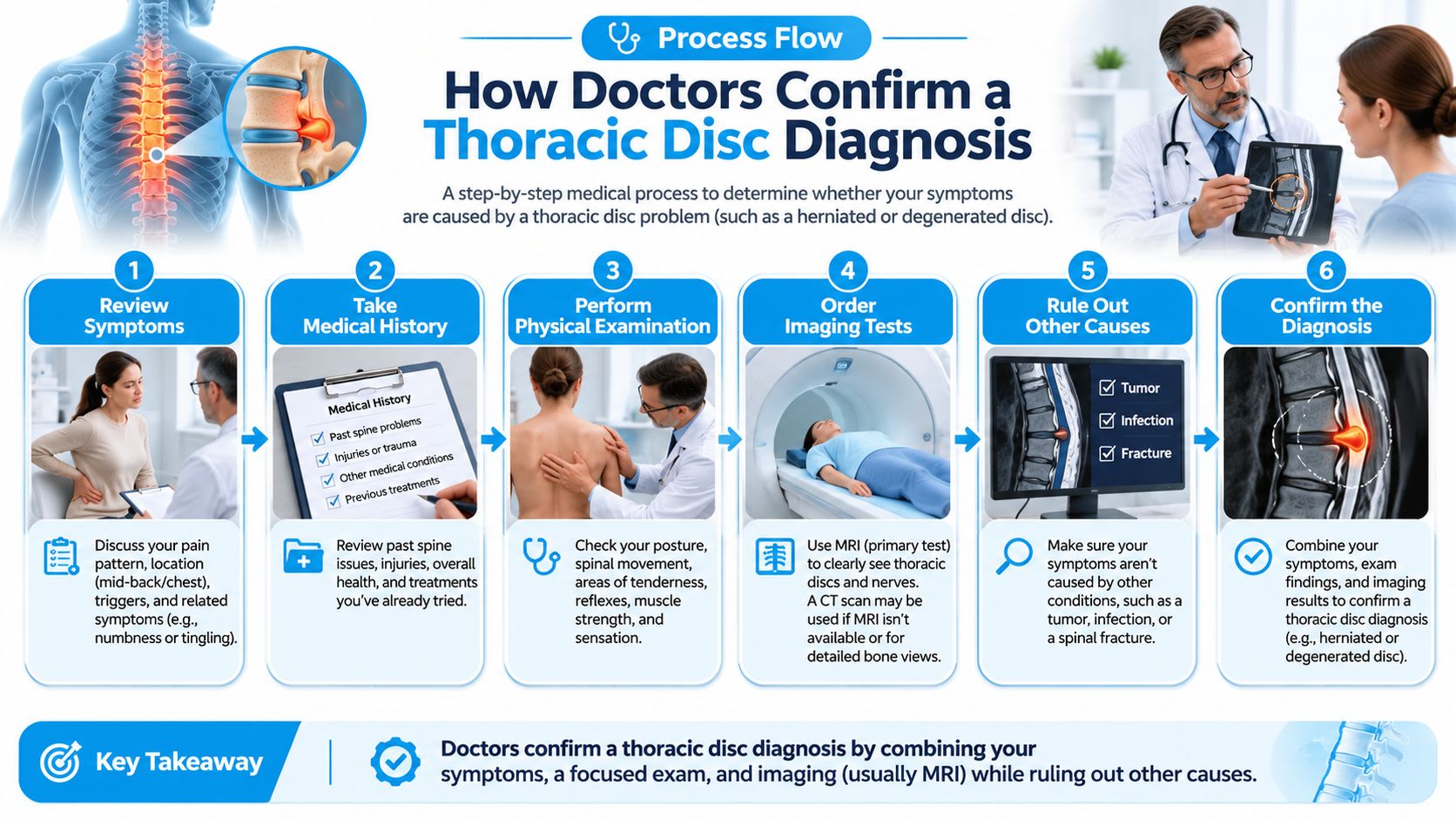
How Doctors Confirm a Thoracic Disc Diagnosis
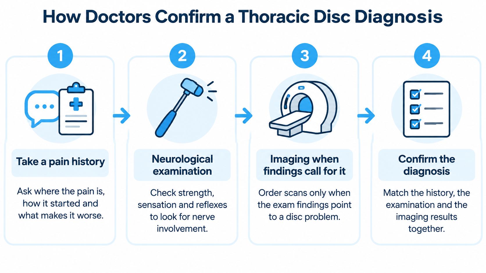
Diagnosis starts with questions and examination, not with selecting a procedure from an MRI report. The clinician needs to decide whether the symptoms are mechanical, nerve-related, spinal-cord-related, or potentially unrelated to the spine.
- Take a pain history and screen for red flags. Explain how symptoms began, where they travel, and what activities change them. Mention relevant medical conditions even if they seem unrelated.
- Check neurological function. Strength, reflexes, sensation, balance, and gait help identify nerve or cord involvement.
- Use standing X-rays when indicated. These help assess alignment, vertebral height, and possible fracture. They aren't automatically necessary for every uncomplicated ache.
- Obtain thoracic MRI when findings warrant it. Persistent symptoms or neurological abnormalities can justify examining the full thoracic segment rather than only a suspected level.
- Match the findings. The location and type of abnormality should plausibly explain the symptoms and examination.
- Reassess over time. New weakness or changing sensation can alter the plan even when earlier imaging seemed reassuring.
The diagnostic process brings those pieces together.
Choose the test that answers the question
Diagnostic tests and what each one answers
| Test | What it shows | When it is ordered |
|---|---|---|
| Neurological examination | Strength, reflexes, sensation, gait and spinal-cord signs | First step in every evaluation |
| Standing X-ray | Alignment, height loss and fracture; selected views assess instability | Trauma, bone-health concern or appropriate baseline assessment |
| MRI of the full thoracic spine | Disc hydration, protrusion, cord compression and endplate change | Persistent symptoms or neurological signs |
| CT or CT myelogram | Bone detail and calcified disc material; myelography outlines compression | When MRI isn't possible or calcification is suspected |
| Blood tests for inflammation or infection | Evidence supporting infection or inflammatory disease | Fever, weight loss or infection risk |
In an MRI study of asymptomatic Japanese volunteers, 46.8% of 94 participants, whose mean age was 48.0 years, had degenerative thoracic findings. A separate longitudinal study described in the same evidence base followed 103 initially healthy volunteers and found that 63.1% progressed by at least one MRI grade over approximately 10 years.
Neither finding establishes that structural change produces pain or disability.
For back or neck nerve-pain symptoms, we recommend starting with an MRI as part of the clinical review. Our lower-back assessment process also starts with MRI, but a thoracic case needs its own localization and examination rather than an automatic lumbar-style diagnosis.
Conventional Care Options and Their Limits
Patients with thoracic disc symptoms and no neurological deficit begin with nonoperative care. The goal is to restore comfortable function while monitoring for changes that require escalation.
A review of thoracic disc pathology and management reports that symptomatic thoracic herniation represents approximately 0.1% to 3% of all spinal disc herniations. It describes conservative management for patients without neurological abnormalities, with surgery generally reserved for myelopathy, significant neurological compromise, or persistent radicular symptoms despite nonoperative treatment.
Conventional thoracic disc treatments compared
| Option | Best for | Main limit |
|---|---|---|
| Activity changes and home exercise | Mechanical mid-back pain with no neurological signs | Needs consistent effort over several weeks |
| Physical therapy for posture and mobility | Stiffness and muscle guarding | Slower relief when pain is severe |
| Short-term anti-inflammatory medicines | Pain flares | Doesn't change disc structure; gastrointestinal and kidney risks with long use |
| Epidural or facet injections | Persistent radicular or facet pain | Relief may be temporary; repeat injections carry their own risks |
| Surgical decompression | Myelopathy or significant neurological compromise | Reserved mainly for cord involvement, progressive deficit, or selected refractory nerve symptoms |
Match the intensity of care to the problem
Activity changes should reduce aggravating loads without turning into prolonged bed rest. For example, raise reading material, support your forearms at a desk, and interrupt long sitting with comfortable movement. A therapist can then select exercises for thoracic mobility, shoulder-blade control, and trunk endurance.
Medication can make movement more manageable during a flare, but it needs a safety check. Tell the prescriber about kidney disease, stomach ulcers, blood thinners, and other medicines before using anti-inflammatory drugs.
Injections serve different purposes depending on the target. An epidural approach aims at nerve-related inflammation, while a facet injection addresses suspected pain from the small joints behind the spine. Neither should be chosen solely because a report contains the word degeneration.
Treatment boundary: Regenerative treatment must not delay decompression when objective spinal-cord compromise requires surgical care.
Conservative approaches mainly address symptoms, movement, and mechanical load rather than reliably restoring disc structure. Surgery is different because it changes anatomy to relieve compression or address instability, but it isn't a general remedy for every mid-back ache.
Our comparison of herniated disc treatment options can support a discussion about alternatives. The useful question is which approach addresses the confirmed source of your limitations, not which sounds most advanced.
Regenerative Stem Cell Options and What Studies Show
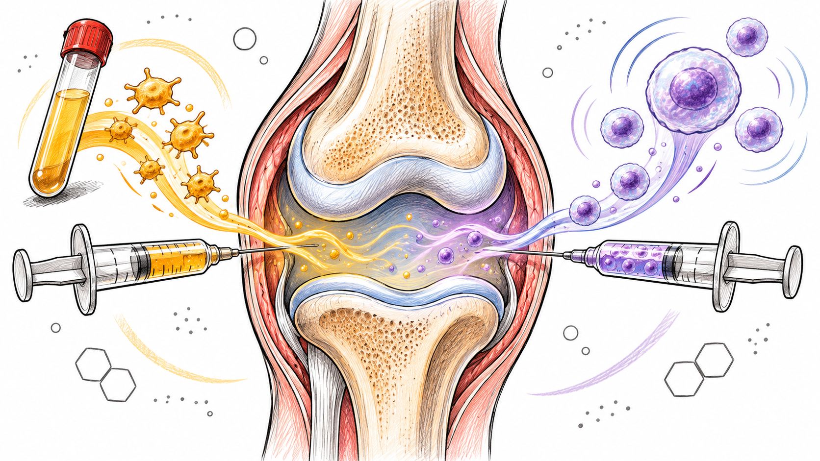
Mesenchymal stem cells, or MSCs, work primarily through the paracrine effect. They release specialized signals, including cytokines, growth factors, and extracellular vesicles, that influence inflammation, tissue survival, vascular responses, and local repair.
For this treatment explanation, the cells aren't understood to differentiate into replacement disc, nerve, cartilage, or bone cells. Their intended role is to guide the healing environment through signaling, not to become a new disc.
Positive clinical findings and their scope
A randomized controlled trial of allogeneic bone-marrow-derived MSCs reported faster and statistically significant improvement in pain-and-function outcomes compared with controls. That supports clinical interest in cell-based disc treatment, but it doesn't establish that every preparation or injection route produces the same result.
A pilot study involving 33 patients with lumbar degenerative disc disease found that 85% of treated patients who completed follow-up MRI had reduced disc-bulge size, with an average reduction of 23%. This was a lumbar study using culture-expanded autologous bone-marrow MSCs, not a dedicated thoracic trial.
A systematic review and meta-analysis of MSC treatment for discogenic pain reported a pooled Visual Analogue Scale reduction of 41.62 points, with a 95% confidence interval of 24.32 to 58.93, p < 0.01, and substantial heterogeneity of I² = 98%. The positive pooled result therefore came with considerable variation between studies.
No dedicated thoracic trial appears in this evidence. Applying these findings to mid-back treatment requires extrapolation, and the studies don't directly validate every clinic's cell product, placement technique, or proposed structural benefit.
What to clarify before treatment
- Delivery route: Intradiscal injection and injections around affected levels aren't interchangeable procedures.
- Risks: Intradiscal procedures can cause infection, bleeding, or nerve irritation. Other routes also require appropriate sterile technique and risk assessment.
- Outcome measures: Track pain, walking, sitting tolerance, daily function, and imaging when clinically appropriate, rather than relying only on a promise of tissue repair.
This use of stem cells is not FDA approved. Dream Body Clinic delivers its treatments under COFEPRIS oversight, Mexico's regulatory counterpart to the FDA; that oversight shouldn't be confused with proof of effectiveness for a particular thoracic condition.
We treat herniated discs without surgery, and herniated-disc care is probably our second-most common treatment area. Our disc degeneration information, lower-back treatment page, and neck-pain treatment page provide context for a case discussion. Outcomes aren't guaranteed, and suitability depends on the diagnosis.
Confirm Your Next Step Before Choosing Treatment
Begin with a doctor who can examine your neurological function and assess any urgent symptoms. Then gather the full thoracic MRI report and images, available X-rays, your medication list, and relevant cancer or infection history.
For a regenerative case review, describe your limitations concretely. Saying that you can't sit through a meal or that pain wraps around a particular rib is more useful than reporting only that your MRI shows degeneration.
Get a thoracic-specific plan
Our middle-back protocol consists of four injections of 25 million cells each, placed around the most affected discs. It addresses herniated discs, ligament or tendon injuries, facet-joint arthritis, and stenosis.
The protocol is designed to support disc rehydration during overnight unloading, restore disc height, rebuild facet-joint cartilage, and reduce stenosis and nerve compression. These are intended treatment goals, not established overnight outcomes or guaranteed structural changes; the clinical studies discussed here don't prove those results for this thoracic protocol.
Before booking, use the complimentary case review and English-speaking phone or WhatsApp coordination to clarify:
- Your target diagnosis: Which disc, joint, or soft-tissue problem best explains your symptoms?
- The proposed procedure: Confirm the cell preparation, placement, dose, and imaging guidance in writing.
- Follow-up: Establish who will reassess you, how improvement will be measured, and what happens if symptoms worsen.
- Alternatives: Ask whether continued rehabilitation or a surgical opinion is more appropriate.
Compare the clinic's descriptions for herniated discs, disc degeneration, and facet-joint problems without assuming that neck or lower-back protocols automatically apply to the thoracic spine.
Choose regenerative care only after urgent causes have been excluded and the examination and imaging support the treatment target.
You don't need to decide from an MRI report alone. A case review should leave you with a clear explanation of what is being treated, why that treatment fits, and what remains uncertain.
Dream Body Clinic offers targeted MSC treatment and case review for thoracic disc and related mid-back conditions. Visit Dream Body Clinic to review the treatment information and submit your imaging and symptom history for a thoracic-specific assessment before arranging travel.