You notice it while trimming your child's toenails, or when a photograph shows your own big toe angling toward the other toes. Perhaps the toe aches in shoes, rubs against its neighbor, or just looks different from the other foot. Big toes that point inwards can describe several unrelated patterns, so the visible position alone doesn't establish a diagnosis.
In a young child, the appearance is often part of intoeing, a rotational alignment pattern arising from the foot, shin, or thigh. In an adult, the same description may refer to hallux valgus, the structural deformity commonly called a bunion, where the great toe drifts laterally while the first metatarsal changes position. Less commonly, an adult has hallux varus, where the hallux deviates medially. These conditions need different examinations and different expectations.
The practical question isn't, “How do I straighten the toe?” It's, “Where does the abnormal rotation or load transfer begin, and is it causing pain or loss of function?” A useful overview of symptom-focused options is bunion foot pain management, but persistent symptoms still deserve an assessment of the whole foot and gait.
Table of Contents
- Understanding Medially Deviated Big Toes
- Pediatric Intoeing Versus Adult Deformities
- The Biomechanics of First Ray Load Transfer
- Evaluating Non-Surgical Management Options
- Surgical Interventions and Iatrogenic Risks
- Regenerative Medicine and the Paracrine Effect
- Navigating Your Treatment Pathway
Understanding Medially Deviated Big Toes
A patient may arrive convinced that the toe itself is the problem. On examination, the toe can be the final visible expression of a mechanical issue that begins elsewhere. In children, the foot may turn inward because of metatarsus adductus, the shin because of internal tibial torsion, or the thigh because of femoral anteversion. In adults, a bunion changes the relationship between the first metatarsal and the hallux, often affecting how the forefoot accepts and transfers force.
That distinction matters because appearance and function don't always match. A child with flexible, painless intoeing may run, climb, and play normally while the alignment gradually improves with growth. An adult with a modest-looking deviation may struggle with footwear, develop pressure over the first metatarsophalangeal joint, or compensate through the ankle, knee, and hip.
The same visual description can hide different anatomy
The phrase “pointing inwards” is imprecise. A clinician asks whether the entire foot progresses inward during walking, whether the big toe angles toward the lesser toes, or whether the toe is deviated medially after previous surgery. Those are separate observations.
For intoeing, the assessment includes the foot progression angle, thigh-foot angle, and hip rotation. These measures help locate the source of the twist. A toe-focused photograph can't provide that information.
Hallux valgus is also more than a sideways toe. Expert descriptions characterize it as a triplanar first-metatarsophalangeal deformity, involving the first ray, the great toe, and surrounding soft tissues. The deformity can alter pressure distribution even when the patient's main complaint is cosmetic.
Practical rule: Treat the symptom that limits walking, footwear, work, or sport, but diagnose the mechanical source before choosing an intervention.
Pain, progressive stiffness, recurrent rubbing, skin breakdown, or difficulty bearing weight should prompt clinical review. The urgency is greater when the alignment is rapidly changing, follows trauma, or occurs with marked asymmetry. A careful history and standing examination usually provide more useful direction than trying to correct the toe from a photograph.
Pediatric Intoeing Versus Adult Deformities
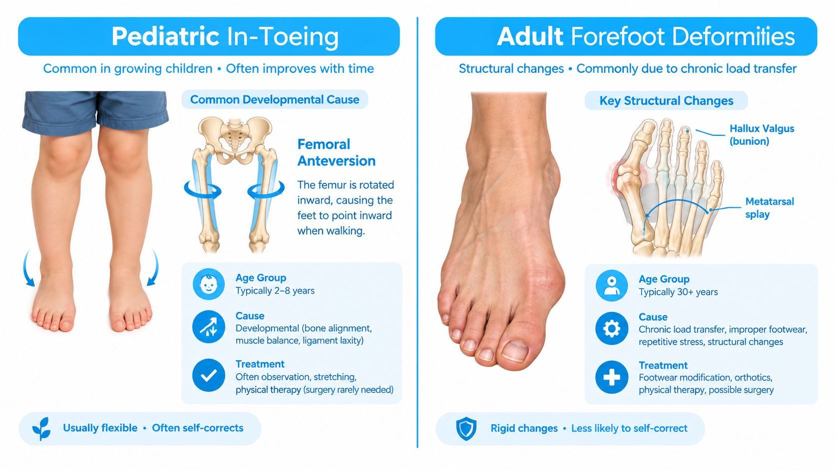
A toddler whose feet turn inward during a walk may have a developmental rotational pattern. An adult whose big toe angles toward the lesser toes usually requires a different assessment. The shared appearance can misdirect treatment unless the clinician identifies which segment is deviating.
In children, intoeing generally arises from metatarsus adductus, internal tibial torsion, or femoral anteversion. The leading cause changes with development. Metatarsus adductus is most common before age one, internal tibial torsion from ages one to four, and femoral anteversion is often recognized after age three, according to OrthoInfo's intoeing guidance.
Growth often improves these patterns without corrective treatment. UCSF clinical information states that metatarsus adductus typically resolves by the first birthday, internal tibial torsion usually resolves by age five, and femoral anteversion commonly improves as the child grows. Observation can therefore be more appropriate than braces, special shoes, or procedures, provided the examination supports a normal developmental course.
Adult forefoot deformity
In adulthood, an inward-looking big toe more often reflects a structural forefoot problem. Hallux valgus involves lateral deviation of the great toe together with altered first-metatarsal alignment. A systematic review and meta-analysis reported a pooled worldwide prevalence of 19% across 186,262,669 participants, rising to 22.7% in people over 60 and measuring 11% in people under 20 (systematic review and meta-analysis). Reported prevalence also varied by region, measuring 21.96% in Asia, 18.35% in Europe, and 3% in Africa.
Those figures describe observed frequency, not a single cause. Foot shape, inherited alignment, footwear, activity, and access to diagnosis may all influence the result. StatPearls reports adult prevalence of approximately 23% among adults aged 18 to 65 and 36% among adults older than 65, with women affected more often (Hallux Valgus, StatPearls).
Hallux varus is a separate adult pattern
Hallux varus is medial deviation of the hallux, opposite to hallux valgus. It is most often associated with previous bunion surgery, particularly when lateral release, medial eminence resection, medial capsular tightening, or correction of the intermetatarsal angle shifts the first MTP joint too far. Its mechanism matters because the problem reflects altered soft-tissue balance and load transfer, not an isolated toe position.
A child with flexible intoeing generally needs developmental observation and rotational examination. An adult may need standing radiographs, footwear assessment, and evaluation of first-ray stability before treatment is selected. The same visual phrase can therefore lead to very different decisions.
The Biomechanics of First Ray Load Transfer
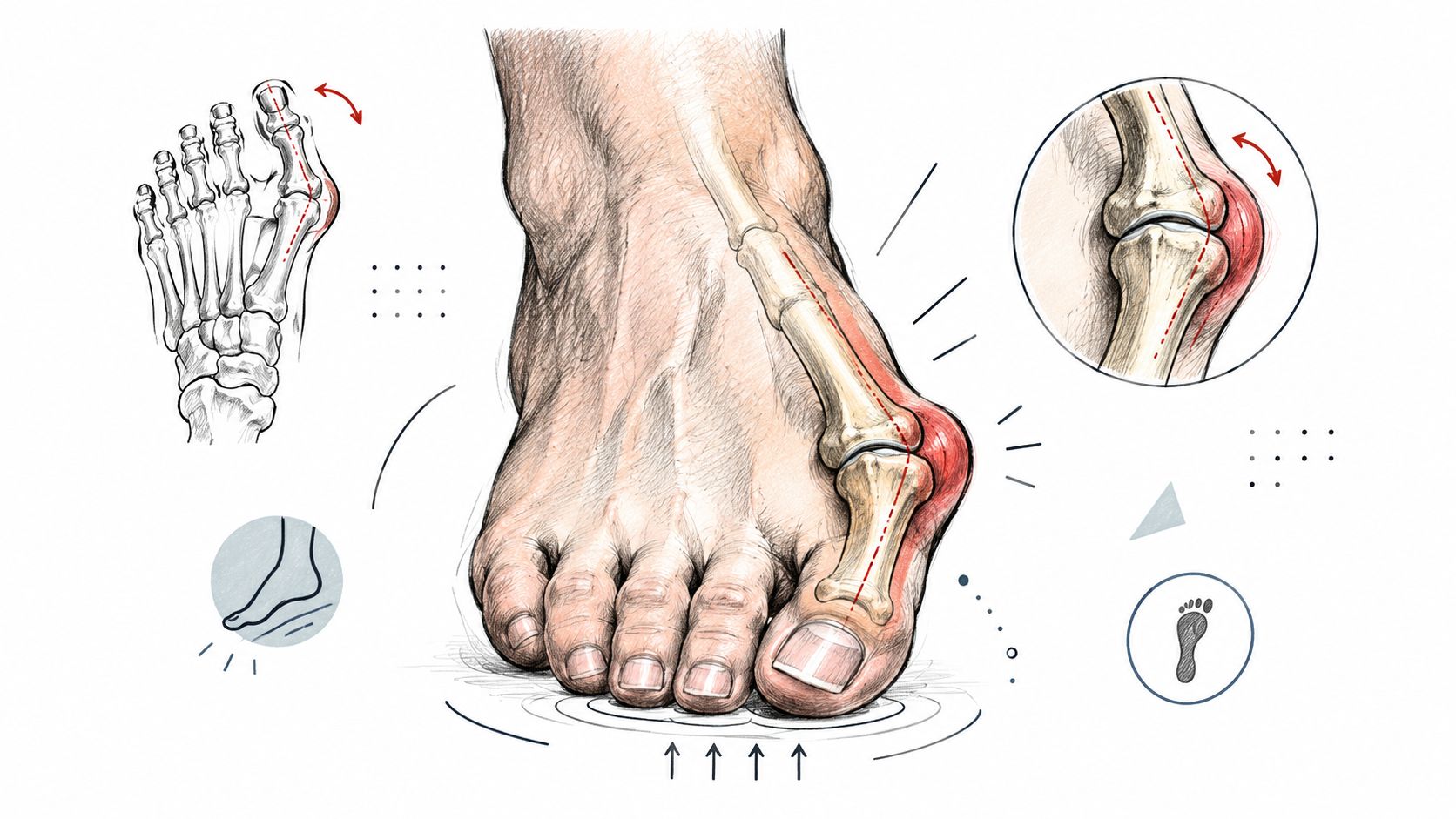
A big toe doesn't work in isolation. During walking, the first ray, made up of the first metatarsal and associated structures, helps the forefoot accept and transfer load. Hallux valgus can disrupt that process through a combination of metatarsal position, hallux deviation and pronation, first-ray hypermobility, ligamentous laxity, metatarsus adductus, pes planus, and equinus contracture.
How a clinician finds the source
A useful assessment follows the load, not just the outline of the toe.
-
Observe standing alignment. Weight-bearing reveals how the arch, heel, first ray, and hallux behave under load. A non-weight-bearing examination can underestimate the deformity.
-
Check first-ray mobility. Excess movement can make a toe splint look helpful while leaving the underlying instability unchanged. The finding may influence whether an orthosis, strengthening plan, or surgery is appropriate.
-
Review gait and footwear. Narrow or high-heeled shoes increase first-metatarsal loading and valgus stress during gait. A shoe that compresses the forefoot can aggravate symptoms without being the original cause.
-
Use standing radiographs when indicated. Imaging helps define bone alignment and joint congruence. It becomes particularly important when pain persists, the deformity progresses, the joint is stiff, or an operation is being considered.
-
Assess the kinetic chain. In children, hip rotation, femoral alignment, tibial rotation, and foot shape help identify intoeing. In adults, ankle motion, pronation, arch behavior, and proximal compensation can change the way the forefoot loads.
A toe-straightening exercise can improve control, but it can't reliably overcome a structural load-transfer failure on its own.
This is also why ankle or lower-limb symptoms may deserve separate evaluation rather than being attributed automatically to the big toe. Patients comparing forefoot complaints with other musculoskeletal concerns may find this clinical discussion of damaged ankle tendons useful as general context, although it isn't a substitute for a foot examination.
A visual demonstration can help patients understand how the first MTP joint participates in alignment and propulsion.
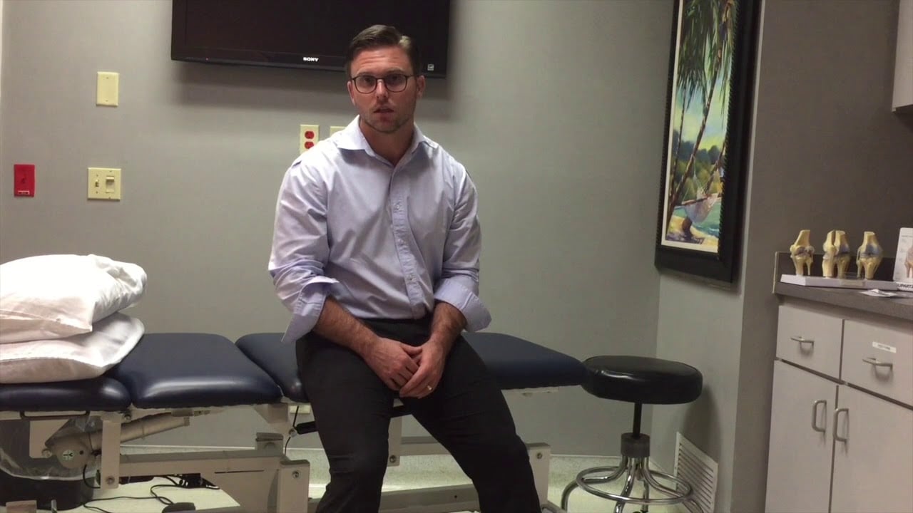
The goal isn't to chase a perfect-looking toe. It's to determine whether the joint can accept load, whether the hallux contributes to propulsion, and whether the surrounding tissues are becoming overloaded.
Evaluating Non-Surgical Management Options
Non-surgical care works best when it's matched to the mechanical problem. Wider footwear may reduce pressure, but it won't stabilize a hypermobile first ray. A splint may temporarily reposition a flexible toe, but it shouldn't be sold as a dependable method for reversing an established structural deformity.
Match the tool to the task
Footwear modification is the simplest starting point for many adults. A deep, wide toe box reduces crowding, while a lower heel can reduce excessive forefoot loading. This approach manages external pressure. It doesn't correct every internal alignment problem.
Orthoses can influence load distribution and arch mechanics. A clinician may choose an off-the-shelf device for mild symptoms or a custom orthosis when pronation, pes planus, or first-ray instability needs more specific control. The fit matters. An insert that creates a new pressure point can make symptoms worse.
Physical therapy should be targeted rather than generic. Programs may address the abductor hallucis, intrinsic foot muscles, calf flexibility, balance, and gait strategy. The purpose is improved muscular control and more favorable loading, not the promise that exercises will remodel a rigid bone deformity.
Gait retraining can be valuable when the patient repeatedly loads the medial or lateral forefoot in a way that provokes symptoms. A therapist may use video feedback, controlled walking drills, and progressive strengthening to build a more efficient pattern.
Recent evidence supports taking this biomechanical approach seriously. A 2025 systematic review found lateral metatarsal loading increased by 40% to 55% in hallux valgus models, while a 2025 meta-analysis found reduced hallux loading on in-shoe sensors (PubMed review). These findings describe altered load transfer, not merely an appearance problem.
What conservative care can and can't do
Conservative management may reduce pain, improve shoe tolerance, support function, and delay escalation. It usually can't guarantee correction of a severe, rigid deformity, and it won't remove a structural bunion just because the patient wears a splint at night.
The most useful plan has a measurable purpose. That might be walking comfortably in ordinary shoes, returning to a sport, reducing pressure over the first MTP joint, or maintaining function while monitoring progression. If symptoms remain limiting despite a well-matched plan, the next step is a specialist review, not an endless cycle of increasingly expensive devices.
Surgical Interventions and Iatrogenic Risks
Persistent pain, loss of function, and mechanical overload can justify surgery after suitable conservative care. The aim is load correction, not a cosmetic photograph. Hallux valgus is a three-dimensional deformity involving the first metatarsal, hallux, sesamoids, joint stability, and the way the patient bears weight.
Operations may combine osteotomy, first-ray realignment, and soft-tissue release or repair. Selection depends on deformity severity, joint condition, mobility, and radiographic findings. A correction that appears convincing on an image may fail if it ignores first-ray hypermobility or the patient's habitual loading pattern.
The risk of correcting too far
Hallux varus shows how an alignment target can create a new mechanical problem. Excessive lateral release, removal of too much of the medial eminence, an overly tight medial capsule, or excessive correction of the intermetatarsal angle can push the first MTP joint into medial deviation. Disruption of the sesamoid apparatus and capsuloligamentous stabilizers may leave splinting unable to restore balance.
Orthopedic reviews associate a notable minority of hallux varus cases with iatrogenic overcorrection (hallux varus surgical review). This is not an individual prediction, but it supports preserving soft tissue and setting an accurate correction target.
Surgical decision: Persistent functional pain and mechanical failure should guide the indication, rather than concern about appearance alone.
Recovery carries its own trade-offs. Bone healing, swelling, limited footwear, stiffness, altered sensation, recurrent deformity, and transfer pain can all affect the result. Ask how the surgeon will assess first-ray stability, which alignment target will be used, how recurrence will be monitored, and how a new imbalance would be managed. A general discussion of postoperative musculoskeletal symptoms, including knee popping after ACL surgery, also demonstrates why recovery planning deserves the same attention as the procedure.
Wound problems can change the treatment priority. Pressure lesions, skin breakdown, and delayed healing around a bunion require direct assessment and protection. The resource bunion wound care expert tips at Vein provides practical wound-care guidance. Managing the wound does not realign the toe, but healthy skin is necessary for safe recovery and sustained weight-bearing.
Regenerative Medicine and the Paracrine Effect
Regenerative medicine addresses a different part of the problem from osteotomy. It may be considered when joint inflammation, tissue stress, or degenerative change contributes to pain, but it doesn't mechanically straighten a rigidly displaced toe. A patient with severe hallux valgus still needs an honest discussion about footwear, orthoses, surgery, and load transfer.
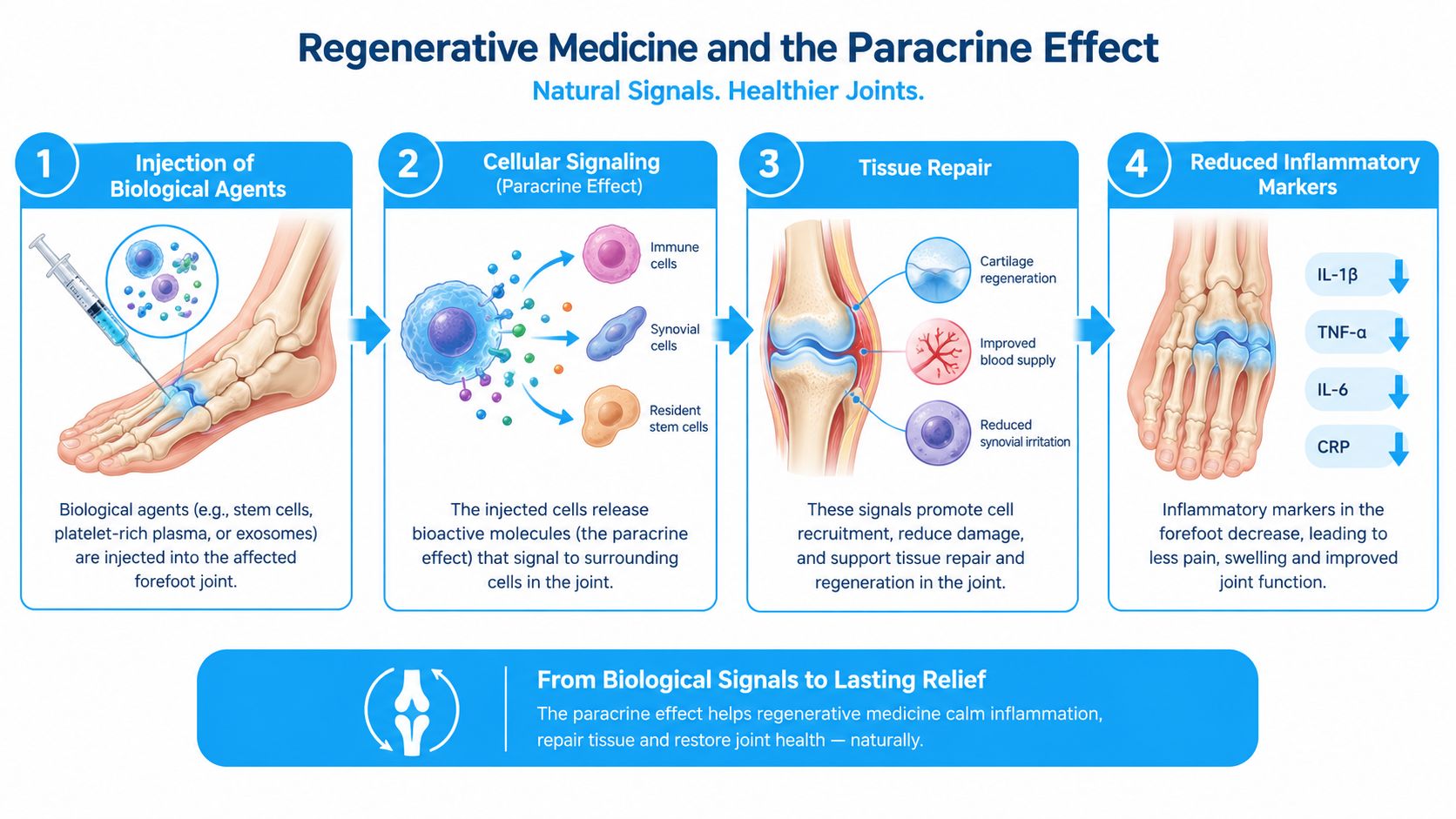
The central biological point is often misunderstood. Mesenchymal stem/stromal cells, or MSCs, don't primarily work by differentiating into replacement cartilage, bone, or other tissue cells. They work mainly through the paracrine effect, sending specialized signals that guide the healing process.
What the signals do
MSCs behave more like a temporary signaling factory than a supply of replacement parts. Reviews describe a “hit-and-run” model in which the cells release cytokines, growth factors, extracellular vesicles, and other soluble factors that influence nearby cells (MSC paracrine mechanism review). These signals can affect immune activity, tissue remodeling, cellular homeostasis, and the local response to injury.
A further review of the MSC secretome describes growth factors, cytokines, chemokines, extracellular matrix components, and metabolic products as contributors to immune modulation and tissue remodeling (MSC secretome review). In practical terms, the aim is to change the local biological environment, not to claim that injected cells become new joint structures.
Biological reality: MSC therapy is a signaling strategy for tissue modulation. It isn't a substitute for correcting every mechanical deformity.
An osteoarthritis-focused review similarly describes MSC activity as mainly involving regulation of inflammation and immune responses through soluble cytokines or exosomes, despite the cells' capacity for chondrogenic differentiation (Frontiers review on MSCs and osteoarthritis). A wound-repair review also concludes that most therapeutic effects are likely related to soluble factors that regulate local cellular responses, including inflammation, angiogenesis, and fibroproliferation (MSC wound-repair review).
For a patient with an irritated first MTP joint, this distinction helps set realistic goals. The potential target is reduced inflammatory signaling and improved tissue homeostasis around a stressed joint. The treatment decision should still depend on imaging, alignment, symptoms, joint status, and whether the dominant problem is biological irritation or mechanical instability. Patients wanting a plain-language explanation of the process can review how stem cell injections work, then discuss whether the proposed therapy addresses their actual diagnosis.
Navigating Your Treatment Pathway
Start with the pattern, not the label. Ask whether the entire foot turns inward during walking, whether the big toe angles toward the lesser toes, or whether the toe deviates toward the inside after surgery. Age, pain, flexibility, progression, and function then narrow the possibilities.
A practical sequence
-
For a child, assess development and function. Flexible, painless intoeing with otherwise normal activity often supports observation. A clinician should review marked asymmetry, pain, limping, functional limitation, or persistence outside the expected developmental pattern.
-
For an adult, examine the whole first ray. Standing alignment, first-ray mobility, footwear, gait, and joint motion matter more than a non-weight-bearing photograph. Standing radiographs may be appropriate when symptoms persist, the deformity progresses, or surgery is under consideration.
-
Use conservative treatment purposefully. Choose footwear, orthoses, strengthening, and gait work according to the load-transfer findings. Track function and pain rather than expecting a device to erase a fixed bony deformity.
-
Escalate when the problem is limiting life. Recurrent skin breakdown, increasing pain, stiffness, difficulty walking, failed conservative care, or a postoperative medial deviation warrants specialist review. A new deformity after surgery deserves particular attention because soft-tissue imbalance can become harder to correct.
-
Consider regenerative care only after diagnosis. MSC-based treatment may be discussed when inflammation or tissue stress is clinically relevant, but patients should ask what biological target is being treated, what mechanical problem remains, and how follow-up will measure progress.
International patients should request a case review based on current imaging, medical history, medications, prior operations, and functional goals. They should also clarify who manages follow-up after returning home, how complications are handled, and whether the proposed treatment is intended to reduce symptoms, preserve joint function, or defer surgery. No injection should be presented as a guaranteed way to straighten a structurally misaligned toe.
Dream Body Clinic offers case reviews and regenerative consultations for orthopedic concerns, including targeted MSC approaches for foot and big-toe conditions, with treatment delivered in Bucerias, Nayarit, Mexico, under its stated COFEPRIS-regulated framework. Visit Dream Body Clinic to submit your records, discuss whether paracrine-based therapy fits your biomechanics, and plan appropriate follow-up before making an international treatment decision.